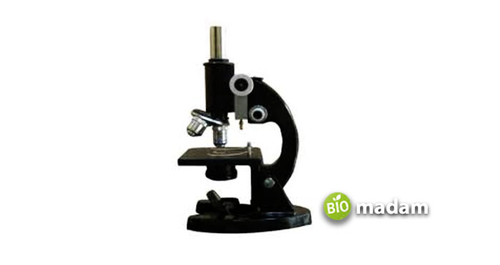
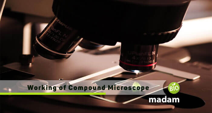
The compound microscope is a type of microscope that is used to magnify those objects which cannot be seen with a simple microscope. Compound microscopes are specially designed for these tiny microorganisms such as bacteria, viruses, etc. In ancient times, an optical instrument was used but it contained a simple lens that does not have much magnifying power, now a compound microscope has an ocular lens and it is used to identify naked eye objects.
Mechanical Components
The tube body of the microscope separates the objective and eyepiece. It is related to the distance between the height of a bench or table talk. It is typically fitted with a turret that rotates and permits the objectives of various powers to be interchanged with the assurance. So, the position of the mage will be maintained. Microscope objectives are used to lessen aberration at the specified tube length. Traditional microscope requires moving the objective of the tube and the eyepiece at the rigid unit.

The specimen is usually placed on a glass slide. The specimen is dipped in a material with an R.I that matches that of the slide and is covered with the thin slide. It allows the glass slide to move across the stage in two directions. So that, the area of a specimen can be explained. This is also suitable for microscopes for students.
As the depth of focus of the objective decreases the accuracy of the movement of the slide increases. The depth of the focus can be lowered by 1 or 2 micrometers which means that mechanical components provide stable motion. In a single mechanical system, incorporating adjustments for the vertical movement is needed for focusing and the lateral motion of the object.
An objective droplet of 160mm of the focal length is adjusted in the lower end of the tube. The objective itself is a maid for the correction of infinite distance.
In some microscopes, the eyepiece is treated as a portion of the zoom lens which permits continuous variation of magnification without the loss of focus. These types of various microscopes are used in industries.
The Illumination System
This system is made to transmit light through a translucent object for vision. It consists of an electric lamp which is a light source and a lens following the condenser. The condenser concentrates the light and focuses the image of light on the plane of the specimen. This technique is called critical illumination.
In Koehler illumination, the entrance pupil of a microscope objective is averaged in the imaging process. The condenser illuminates the specimen against a black background. The use of color filters enclosing years of the nineteenth century by British microscopist Julius Reinberg and now known as Reinberg illumination.
Objective of Compound Microscope
The focal length of the objective is inversely proportioned to the magnification. Focal length increases as the magnification decreases. An objective that uses special glasses made up of crystalline material is called apochromatic. And they were first produced by abbe in 1870.
The correction of spherical aberration is achieved when normal optical glasses are used for the lens element. Conventional objectives do not produce a flat image surface. Field curvature is a problem for imaging systems. However, special objectives with flat field lenses have been designed for these systems.
High Power Objectives
The highest power microscope objective available is the immersion objective. For using this type of objective a drop of oil is placed between the object on the slide and the objective. Water immersion lenses are available.
Depth of Focus
The highest possible resolution can be obtained along the axis of microscope optics. It banned the focusing requirement of the objective. However, contrary to compound microscopes, electron microscopes have a 250x resolution power than the former.
The Eyepiece

It is used to visualize the relating images. The observer places the eyepiece at the pupil of the point. In most cases, 1cm is desirable.
Image Formation
In the absence of aberration, geometric rays form an image point. In the presence of aberrations, it is indicated as the indistinctive point. The focal length and magnification are attained by the aperture of the pupil of the eye.
In 1873, ERNST ABBE suggested that an object in the focal plane of the microscope is illuminated by the light from the condenser. He showed that the greater the number of waves, the finer the image. The diffracted light would be 0.55. The wavelength of light is shortened when it is propagated through a Dense Medium.
The index of refraction is multiplied by N.A of 1.4. Leeuwenhoek shows that the capacity for resolving fibrils in single lenses is 0.7 mm.
Conclusion
The compound microscope has very high magnifying power so it is capable for to be used for seeing very tiny objects in detail it can also be used to see small tissues yeasts or bacteria. Due to the microscope’s huge importance in the educational sector, it is also used in School, College & University labs. Biomedical and other labs are incomplete without a microscope. It is also being used in the process to see the small electrons and is also used in diagnostic labs. As I can see it is also very useful in understanding the structures of tissues, so it is very important in the medical field and also in physics and many other fields.

Jeannie has achieved her Master’s degree in science and technology and is further pursuing a Ph.D. She desires to provide you the validated knowledge about science, technology, and the environment through writing articles.

