All living organisms come into being and reproduce through specific cell divisions. The eukaryotic cells, being multicellular, produce new cells following mitosis and meiosis. Are you familiar with the difference between these two? Mitosis and meiosis and similar and distinct at the same time. The process works through dividing a diploid cell, which converts to daughter cells.
An actively dividing cell undergoes a series of stages known as the Cell Cycle. These usually include Interphase [Gap phase (G1 phase), G2 phase, and Synthetic phase (S phase)] in which the cell prepares itself to duplicate and the Mitotic phase.
Mitosis
Mitosis is cell division that produces daughter cells with the same genetic material and approximately an equal number of organelles and cytoplasm as the parent cells. The hereditary substance equally doubles into daughter nuclei. An organism starts with mitosis soon after receiving particular signals, such as the presence of growth factors. Mitosis is responsible for growing the somatic cells, for instance, blood cells, epithelial tissues, skin cells, etc. This mechanism helps replace the dead or decayed cells out of the body.
Types of Mitosis in Eukaryotes
Eukaryotic cells have two types of mitosis:
Open Mitosis
Open mitosis occurs in animals where the nuclear envelope breaks down to separate the chromosomes.
Closed Mitosis
Closed mitosis takes place in the fungal nucleus, promoting the segregation of chromosomes.
Stages of Mitosis
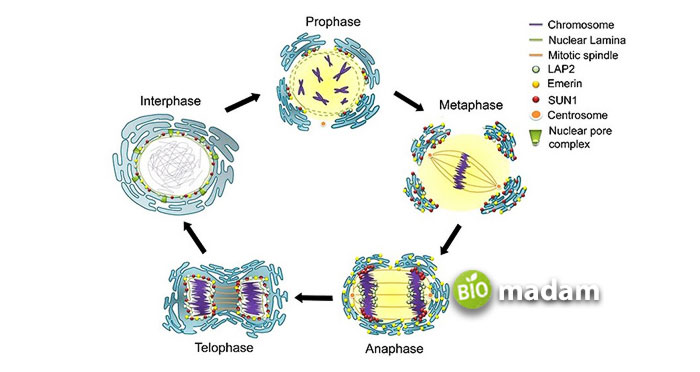
A cell divides through mitosis concerning the following four stages:
- Prophase
- Metaphase
- Anaphase
- Telophase
Preprophase
This phase is an additional step that only takes place in plant cells. The preprophase starts when the nucleus of highly vacuolated plants migrates to the center of a cell. The centrosome disappears, which gives rise to the ring of actin filaments along with microtubules. In other words, the coordinating center of microtubules eliminates division.
Prophase
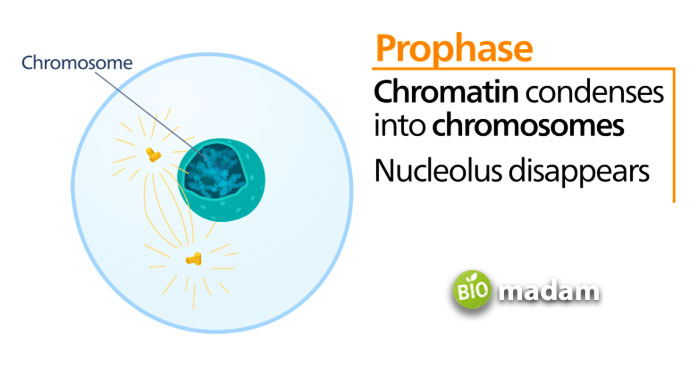
It is the first and foremost stage of mitosis, where the nucleolus disappears. The nucleus contains a material called chromatin which condenses into a rigid structure called a chromosome. These can be of many types that further help break down the nuclear envelope. Besides, the spindle fibers are formed at the opposite ends of a cell that will eventually help it divide into daughter cells. The prophase of mitosis takes much less time than the prophase I of meiosis.
Metaphase
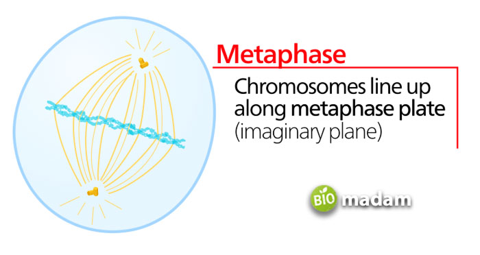
Some experts say the nuclear envelope disappears during the prophase, and some say it’s a prometaphase stage of the cell. Every chromosome has a centromere called the kinetochore. When spindle fibers form, they bind to kinetochores to produce a metaphase plate. Similarly, the non-kinetochore fibers (not attached to the centromere of chromosomes) make a bond with fibers of their type. The chromosomes align with the metaphase plate of the spindle apparatus during the metaphase.
Anaphase
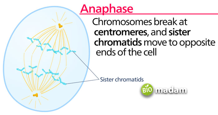
During anaphase, each pair of chromosomes is separated into two identical chromosomes by the mitotic spindle and pulled to opposite cell poles. It ensures each daughter cell receives an identical set of chromosomes, and these chromosomes are segregated to reform the nucleus for further functions.
Telophase
Telophase is the final stage of mitosis that is reverse of prophase. During this stage, reappearance and enlargement of nucleolus and dissolution of the kinetochore microtubules occur. As the nuclear envelopes re-form, the chromosomes begin to de-condense and diffuse more to form chromatin. A nuclear envelope starts forming around the chromosomes. Telophase ends at a cytokinesis process either through mitosis or meiosis.
Significance of Mitosis
Mitosis plays a significant role in all living entities with numerous benefits. These are:
Genetic Stability
We all know mitosis maintains the number of chromosomes in both parent and daughter cells, which helps in genetic stability. All the cells formed would be completely identical to each other, thus managing the population.
Development and Growth
Mitosis helps increase the number of cells in a living organism. For example, the embryo grows from the fertilization of gametes by mitosis, and this growth continues throughout an organism’s life.
Replacement and Regeneration of New Cells
The replacement and regeneration of the cells is an essential function of mitosis that helps repair the damaged tissue or replace the worn-out cells. An example of this is healing a cut or a broken bone, replacing the old dead cells like RBC. These cells die after 120 days, and new cells form through mitosis. Another famous example is the growth of the lost arm of a starfish via mitosis.
Asexual Reproduction
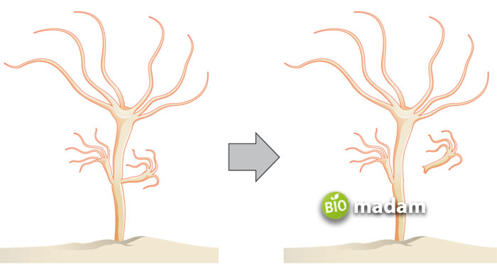
Some organism reproduces through asexual method rather than sexual reproduction. This method promotes genetically identical offspring. This type of reproduction follows mitosis division. For example, hydra uses mitosis and reproduces by budding or binary fission. Yeast cells also multiply through budding and reproduction by fragmentation, as in a Planarian.
| Differences | Mitosis | Meiosis |
|---|---|---|
| Mode of replication | Asexual reproduction | Sexual reproduction |
| Occurrence | Somatic cells of all organisms Absent in viruses | Germ cells of animals, plants, and fungi |
| No. of divisions | One | Two |
| Crossing over | Absent | Present |
| Chromosomes number | Remains the same | Reduced by half |
| No. of daughter cells | 2 diploid cells | 4 haploid cells |
| Chiasmata | Absent | Present in prophase I and metaphase I |
| Exchange of Segments | Occur in prophase | Occur in prophase I |
Meiosis
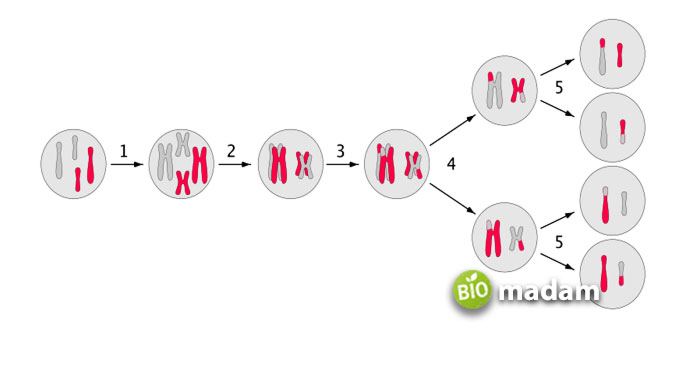
Another type of cell division is meiosis, which leads to daughter cells having half the number of chromosomes as the original ones. Meiosis reduces the overall chromosome numbers from 46 to 23. A single living cell divides twice to produce four offspring cells with one-half chromosomes and genetic information.
The principal function of meiosis is to produce spores or gametes (sex cells) through the process called spermatogenesis and oogenesis. These respectively produce sperms and eggs in mammals. These finally contain a haploid number of chromosomes.
Types of Meiosis in Eukaryotes
Eukaryotic cells undergo two types of meiosis:
- Meiosis I
- Meiosis II
Meiosis I
The first division, called the reduction division of meiosis, is in which homologous chromosomes start separating. The four primary stages of meiosis-I are as follow:
- Prophase I
- Metaphase I
- Anaphase I
- Telophase I
Prophase I
Prophase I is the most prolonged phase of meiosis, where chromatin condenses to form homologous chromosomes. Here the nuclear membranes begin to dissolve. The homologous chromosomes form bivalents, and synapsis between these materials takes place. The point where two non-sister chromatids from synapsis are called Chiasmata. It is the region where crossing over (exchange of genetic material between non-sister chromatids) occurs. All these events manage the genetic recombination through the following five sub-phases:
Leptotene
Chromosomes start condensing and distinguishing in the leptotene phase of prophase I. These are attached to the nuclear membrane of the cell via their telomeres.
Zygotene
A synaptonemal complex produces between the homologous chromosomes due to the synapsis process. All this happens in the zygotene phase.
Pachytene
In pachytene, homologous recombination, including chromosomal crossover, takes place between non-sister chromatids.
Diplotene
It is an extensive resting phase of prophase I where homologous pairs remain attached at the chiasmata, and the synaptonemal complex disappears.
Diakinesis
The last stage of prophase-I is the diakinesis, where chromosomes fully condense and characterize by chiasmata termination.
Metaphase I
During metaphase I, the bivalents arrange themselves at the opposite poles where microtubules are present. A metaphase plate helps the aligning of these bivalents in metaphase I.
Anaphase I
Microtubules begin to shorten, and centrioles help spindle fibers separate the homologous pairs. These chromosomes move to opposite poles of the cell in a process known as disjunction. The sister chromatids of each chromosome always stay connected in anaphase I, called the sister chromatids.
Telophase I
This phase is the last phase of meiosis I, right after anaphase I. In it, the chromosomes finish moving to the opposite ends of a cell and finally de-condenses. The chromatin materials are again formed, and the nuclear membrane starts appearing. Telophase I also helps in cytokinesis, further forming two non-identical haploid daughter cells.
Meiosis II
The two new cells produced by meiosis-I now enter a second meiotic division called meiosis-II. It may begin with interkinesis or interphase-II which is different from interphase-I. This particular phase doesn’t include the S phase. Similar to meiosis-I, meiosis-II also has four sub-phases, such as:
- Prophase II
- Metaphase II
- Anaphase II
- Telophase II
Prophase II
Prophase immediately sets off after cytokinesis when the cytoplasmic division forms daughter cells. It is pretty similar to the prophase of mitosis. During prophase II the chromosomes begin to condense, and centrioles duplicate. The two pairs of centrioles separate into two centrosomes, and the nuclear membrane starts disappearing. Similarly, other organelles like Golgi apparatus and ER complex also start eliminating.
Metaphase II
All chromosomes connect to the opposite centrioles at the kinetochores of sister chromatids through microtubules. These align in an almost straight line, in between the cell, through a metaphase plate.
Anaphase II
The sister chromatids are pulled apart by the kinetochore microtubules, simultaneously splitting each chromosome’s centromere. This way, these sister chromatids are pulled away towards the opposing poles.
Telophase II
The fourth and last step of meiosis II is telophase II. Here, the chromosomes reach the opposite poles and de-condenses there. The lost nuclear envelope reappears around the chromosomes, and cytokinesis occurs. It leads to the formation of four haploid daughter cells followed by meiosis-I and all previous steps of meiosis II.
Significance of Meiosis
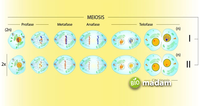
Meiosis is equally essential as mitosis, playing a significant role in all living entities. These are:
Formation of Sex/Gamete Cells
The principal function of meiosis is to form spores/gametes (sperm and eggs in mammals) that have the haploid (n) number of chromosomes and required for sexual reproduction.
Maintenance of Chromosomal Numbers
Meiosis maintains the constant number of chromosomes in sexually reproducing organisms with the help of meiosis I and II during gametogenesis.
Introduces Variations in Generations
In meiosis, the reshuffling of chromosomes happens through a particular procedure called crossing over. Thus, this phenomenon produces a new combination of traits and variations in offspring cells.
Mutations
Chromosomal and genomic mutations occur by irregularities in cell division by meiosis. Some of these mutations are useful to the organism and carried on by a natural selection, for example, lactose tolerance.
Which Process is More Worthy – Mitosis of Meiosis?
Both cell division processes are significant in maintaining a living organism. If mitosis helps in genetic stability, meiosis provides variations and different traits for the continuity of life. Hence, grabbing the difference between mitosis and meiosis is more critical than comprehending which one is more worthy.

Hello, I would like to introduce myself to you! I am Chelsea Rogers, an experienced blog writer for science articles, holding an MPhil degree. My enthusiasm to grab the best knowledge, let it relate to botany, zoology, or any other science branch. Read my articles & let me wait for your words s in the comment section.

