Golgi apparatus or Golgi bodies are a bundle of flattened sacs within the cell that contribute to many cellular functions. Major Golgi apparatus functions include sorting and transporting different types of proteins to send them to other parts of the cell. It also contributes to modifying these proteins before packaging them into vesicles. The Golgi apparatus is found in the cytosol of cytoplasm near the endoplasmic reticulum, next to the nucleus that has a variety of functions. Most cells contain only one Golgi apparatus, yet plant cells may have hundreds of them.
History of Golgi Bodies
The Italian cytologist ‘Camillo Golgi’ observed the organelle in 1897. He established a staining technique to observe nervous tissues that he called “reazione nera,” which means “black reaction.” It led to the discovery of the Golgi bodies, obtaining the name Golgi stain. He fixed the nervous tissues with potassium dichromate and saturated them with silver nitrate. Golgi bodies took on the stain while he was observing the neuron cells, and Camillo Golgi named them “internal reticular apparatus.” The word spread among scientists, but it actually got confirmed in 1950s when appropriately observed under the electron microscope. Unlike a compound microscope that uses a beam of light, an electron microscope utilizes electronic beams to help understand the exact structure of the organelle.
Golgi Bodies Location
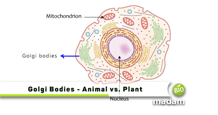
The Golgi bodies are located between the cell membrane (often confused with plasma membrane), and the endoplasmic reticulum near the nucleus. Typically the Golgi apparatus looks like an extension of the ER due to its smooth appearance, like the smooth endoplasmic reticulum, as observed by the scientists who initially examined the Golgi apparatus. Yet, as we mentioned above, Golgi bodies are distinct cell organelles responsible for specific functions.
Golgi Bodies Structure
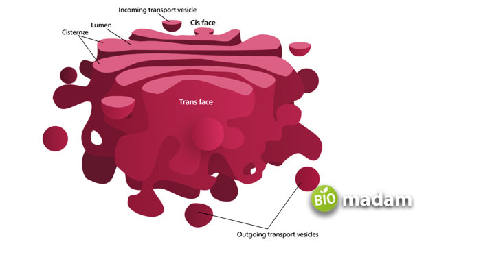
The Golgi bodies appear as a stacking of 5 to 8 compartments curved on one side that may look like a cup. These compartments are known as cisternae. They are disk-shaped, flattened pouches on top of each other to give a stacked look. The cisternae join through matrix proteins, whereas cytoplasmic microtubules hold the complete structure. While commonly, every cell has around 8 cisternae, some protists have more than 60 cisternae; mammalian cells contain between 40 and 100 stacks.
The Golgi bodies comprise a trans and a cis face, with the cis side membranes being thinner than the trans side. The structure is polarized and contains three basic compartments between the trans face and the cis face. The third segment is the medial face comprising the central layers of cisternae. The sides of the Golgi bodies are unique from each other and contain specific enzymes for different functions. They collectively contribute to protein and lipid sorting, which are received by the cis side and released by the trans side.

Golgi Apparatus in Animal Cell vs. Plant Cell
In animal cells, the Golgi apparatus is the main site for protein transportation to other sites within the cell. After modification, the Golgi apparatus sorts and assembles proteins and lipids into vesicles. It provides specific areas within the apparatus where reactions may occur in optimum conditions. The Golgi apparatus also performs protein glycosylation and polysaccharide metabolism within the eukaryotic cell.
However, the transport of materials through Golgi bodies is slightly different in the plant cell. The Golgi apparatus in the plant cell acts as the site for synthesizing polysaccharide molecules required to form the cell wall. Pectin and hemicellulose are produced in the Golgi apparatus to produce the outer membrane of the cell. Thus, plant cells may also have more Golgi bodies than animal cells. Moreover, the absence of lysosomes in the plant cells replaces the function with the vacuole. Thus, in certain situations, you may call the vacuole as the lysosome of a plant cell. The content from the Golgi apparatus eventually moves to the vacuole to fuse with it.
The Bottom Line
The Golgi apparatus is an important part of the eukaryotic cell, often confused with the endoplasmic reticulum. It is present near the ER and the nucleus within the cell. This organelle is mainly involved in modifying and packaging substances that must be transported to other parts of the cell. They contribute to various anatomical and physiological roles in the body. Proteins undergo glycosylation while it metabolizes lipids and polysaccharides.
Moreover, it plays a primary role in the sorting and transporting of proteins throughout the cell. Yet, the function of the Golgi apparatus in plants is different from that of animals. Plants do not have lysosomes. Thus, the contents from the Golgi bodies travel to the vacuole and fuse with it.
FAQs
What is Golgi Apparatus and its function?
Golgi apparatus, also known as Golgi bodies or Golgi complex, is a set of flattened stacks located near the endoplasmic reticulum. It plays a major role in modifying and packaging proteins and lipids before transportation. It also sorts and assembles the proteins before packaging them into suitable vesicles.
What are the 3 main functions of the Golgi apparatus?
Protein transportation, glycosylation, and lipid and polysaccharide metabolism are the three main functions of the Golgi apparatus. It also works to sort and assemble proteins and lipids in the body contributing to the production of different structures within the body.
Why is it Called Golgi Apparatus?
The Golgi apparatus is named after the scientist Camillo Golgi who first identified the structure in neuron cells. They seemed like the endoplasmic reticulum due to their smooth surface and layers. These structures took the stain of potassium dichromate that Camillo Golgi prepared to study brain cells.

Hello, I would like to introduce myself to you! I am Chelsea Rogers, an experienced blog writer for science articles, holding an MPhil degree. My enthusiasm to grab the best knowledge, let it relate to botany, zoology, or any other science branch. Read my articles & let me wait for your words s in the comment section.

