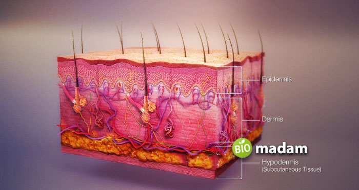
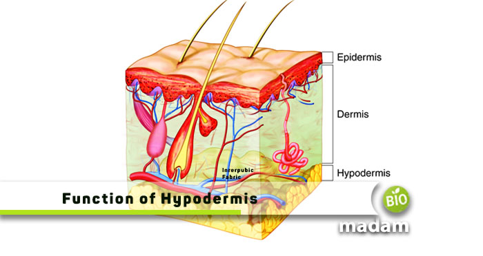
The skin is the largest organ of the human anatomy in size. It protects the underlying skeleton from external elements. When hurt, the fat cells in the deepest layer of skin help cushion the blow and keep your vital organs, like kidneys, stomach, intestines, and liver, safe for functioning. The hypodermis is an essential component of healthy skin; without it, the skin would be thin and fragile, unable to perform its necessary tasks. It is one of the three layers of human skin, along with the epidermis (outer layer) and dermis (middle layer).
By the end of this article, you will understand the function of hypodermis and its critical role in health.
What is Hypodermis?
The word comprises the Greek prefix hypo, meaning “beneath,” and the Latin word dermis, meaning “skin.” Hypodermis is also known as the subcutaneous tissue.
The hypodermis is a layer of connective tissue beneath the dermis. Connective tissues and adipose are the primary components of the hypodermis, the deepest layer of human skin. Fibrous proteins like collagen and elastin comprise the connective tissues, which play a crucial role in the hypodermis’s part of supporting the skin’s upper layers.
Functions of Hypodermis
The hypodermis functions as an anchor for the skin to attach to underlying structures like muscle and bone. The elastic fibers and areolar connective tissue make up this pliable tissue. The hypodermis’s primary functions are the following:
Thermal Regulation
The hypodermis contains a layer of adipose tissue, also known as fat cells, which serve as an insulating layer to help regulate body temperature. Fat cells act as a barrier against heat loss, which helps to maintain body heat during colder temperatures.
Energy Storage
Adipose tissue in the hypodermis serves as a site for energy storage in the form of triglycerides. The body can store excess energy through fat cells in the hypodermis and release this energy as needed to fuel bodily functions.
Acts as a Shock Absorber
Below the hypodermis lies the muscles, bones, tissues, and organs, so this layer naturally acts as a shock absorber. This function helps minimize injury risks from any sort of pressure or external trauma.
Mechanical Protection
The hypodermis provides a cushioning effect that protects underlying tissues from mechanical damage. Areas of the body, the palms and the soles of the feet, which experience constant mechanical stress, depend on this characteristic for proper functioning.
Functions in Wound Healing
We will discuss the structure of hypodermis later, having a proper lymphatic system. This immune cell system within the skin layer is involved in the body’s defense against infections and skin diseases. These cells help the skin fight pathogens entering and promote healing in case the skin is breached.
Nerve and Blood Vessel Support
The hypodermis contains a network of blood vessels that supply nutrients and oxygen to the upper skin layers and the underlying tissues. It also has nerve endings that provide sensory information about touch, pressure, and temperature.

Causes the Release of Hormones
The hypodermic adipose tissue produces leptin, which regulates energy intake and expenditure. It transmits signals to the brain to stop you from overeating.
Promotes Sensory Functions
The hypodermis functions to detect pressure and temperature changes as it has sensory receptors attached. However, these receptors are lesser here than in the dermis.
Holds Muscles and Bones Together
One of the critical functions of hypodermis is to connect the skin to the bones and muscles beneath it. This connection is achieved through fibrous connective tissue strands extending from the dermis to the hypodermis and attaching to the underlying structures. This anchoring of the skin to the deeper tissues helps maintain its shape and position and prevents it from sliding around excessively.
Aesthetic Appearance
The hypodermis’s thickness and distribution can affect the skin’s appearance. The skin appears fuller and more rounded in areas where the hypodermis is thicker, such as the cheeks and buttocks. The skin appears more taut and smooth in areas where the hypodermis is thinner, such as the hands and feet.
Anatomy of the Hypodermis
Hypodermis has a unique anatomy, being the innermost layer of the skin. That being said, its coloration depends on how much carotene (a pigment) is present inside. Based on pigment stimulation, a hypodermis layer can be dark or light yellow. Let’s get to knowing where it is actually located.
Where is the Hypodermis Located?
As the Greek word “hypo” means beneath, the hypodermis is the bottom layer of the skin. It is found deep below the epidermis and dermis layers. In most people, around 80-90% of fat is subcutaneous, which varies from person to person. It is different and more in women than in men that indirectly shapes them. Now that you know the prime location of hypodermis, do you wonder about its structure?
What Major Structure Makes up the Hypodermis?
The hypodermis’s structure differs with differences in body weight. The hypodermis on eyelids and external genitalia are the thinnest on your body, measuring less than 1 millimeter. The thickest areas are the belly and buttocks, where they can measure more than 3 centimeters. Hypodermis is observed to be the thickest in the buttocks, thighs, and hips of women and the shoulders and bellies of men.
A single layer of hypodermis includes different structures, including:
- Adipose Tissues: These are the fatty tissues that make up most of this subcutaneous layer and consist of adipocytes. Adipose cells further cluster into structures called lobules.
- Connective Tissues: Collagen and elastin protein make up the connective tissues in the hypodermis layer, giving skin strength and flexibility. These tissues partition these lobules by thin fibrous septa, providing structural support to this skin layer.
- Vessels and Nerves: The hypodermis is the layer of skin beneath the dermis permeable to the blood vessels, lymphatic vessels, and nerves supplying the skin and underlying tissues. All the blood vessels here dilate to keep the body cool, whereas lymphatic vessels play a role in transporting waste products away from the skin.
- Macrophages: These white blood cells play a significant role in innate and adaptive immunity. Macrophages keep the body free of harmful bacteria and viruses.
- Bursa: It is another slippery and fluid-filled sac in your body, found explicitly in the hypodermis. Bursa acts as a lubricant and a cushion for all skin layers.
- Hair Follicles: These unique hair-like structures are actually part of the epidermis and dermis that fold to extend into the hypodermis part of the skin. It is the point where the hair starts growing.

The Bottom Line
The skin is the largest organ of the body, also known as the dermis. It comprises three layers: epidermis, dermis, and hypodermis. The hypodermis is the innermost layer of the skin. It may differ from one person to another and also varies within different body regions. The main functions of hypodermis include thermal regulation, mechanical protection, energy storage, holding muscle and bone together, releasing hormones, and aesthetic appearance. It also helps protect against antigens and pathogens.
FAQs
What is the role of the hypodermis in maintaining body temperature?
The hypodermis maintains the body temperature by storing fats and forming a cushion on the surfaces underneath. Thus, it offers insulation from cold temperatures.
What tissue is hypodermis?
The hypodermis is made of subcutaneous tissue, also known as the superficial fascia. It is the layer of tissue present underneath the skin. Hypodermis comes from the Greek hypoderm, which means “beneath the skin,” indicating the deepest layer of the skin.
Why is the hypodermis a shock absorber?
Besides cushioning and maintaining temperature, the hypodermis also acts as a shock absorber. This characteristic of the hypodermis is attributed to the presence of adipose tissues that store fats.
Does hypodermis contain blood vessels?
The hypodermis contains a rich supply of blood vessels and provides them to the dermal layer for nutrition.
What cell types are in the hypodermis?
The hypodermis contains adipose cells, fibroblasts, and macrophages. The hypodermis is not derived from the mesoderm’s dermatome region compared to the dermis.
Why is hypodermis not part of the skin?
Hypodermis is one of the three layers of the skin but is not considered a part of the true skin. Its role is more prominent in connecting the skin to muscles and bones.
Does the hypodermis have collagen?
The hypodermis contains fats and collagen. The fats provide cushioning, while collagen provides a definite shape to the layer and keeps it in form.

Hello, I would like to introduce myself to you! I am Chelsea Rogers, an experienced blog writer for science articles, holding an MPhil degree. My enthusiasm to grab the best knowledge, let it relate to botany, zoology, or any other science branch. Read my articles & let me wait for your words s in the comment section.

