Stethoscopes have made their way onto doctors’ necks in all parts of the world. So, it must be something very important that doctors always keep with them, even though, they can check the pulse using their hands as well. Then, what’s the need to keep their stethoscopes all the time?
Different types of stethoscopes have multiple medical applications that people are not typically aware of. You see the doctor using the stethoscope to study your heartbeat and chest movement and prescribe medicine. Is it that simple?
Let’s talk about Stethoscopes and their different uses to help you learn them better.
What do Stethoscopes Do?
Stethoscopes are medical tools used to study the sounds produced within the body, typically listening to heartbeat and chest movements. The first design featured a wooden instrument that was modified to make the stethoscopes we see today. The two flexible tubes have earpieces at one end and a chest piece or head piece at the other. The headpieces can be of various types depending on the need. A bell-shaped open-ended chest piece with a flat shape allows you to study the heart sounds better, as it is more suitable for transmitting low-pitched sounds and higher frequencies at the same time. Doctors can change the function of these dual stethoscopes per need.
Thus, stethoscopes help you observe and analyze sounds in different parts of the body. Besides their most common application in examining the heartbeat for a general checkup, they are used to measure blood pressure, understand recovery from surgery, check bowel sounds, etc. Keep reading to learn about these uses of stethoscopes in detail.
Uses of Stethoscope
Heart Auscultation
The phenomenon of listening to body sounds using a stethoscope is known as auscultation and the most common use of a stethoscope is heart auscultation. Doctors use stethoscopes to listen to your heartbeat and study the condition of your heart. Most patients sit on a chair while doctors examine their heartbeat. Sometimes, the doctor may ask you to lay at a 45-degree angle or turn to your left side. They place the stethoscope on your chest to listen to your heart and move around the stethoscope to study all areas in your upper left chest. They notice the heart rate, the length of each heartbeat, the intensity, and the frequency of your heart sounds.
Lung Sounds
The second most common type of auscultation recorded by the stethoscope is lung sounds to diagnose the type of respiratory disease. The person under examination is asked to sit straight and breathe deeply through their mouth. The doctor then places the stethoscope on the chest and moves it to different points to study both parts of lungs. Starting from the top towards the end of the lungs, they record and compare the sounds of the lungs. The same process is repeated on your back to examine breathing patterns and air movement through the respiratory system and check if there is any breathing issue. The doctor will study each cycle and your breathing rate. Observing the sounds of your lung by examining them from the back gives them information about any difference in those sounds. The doctor will measure the quality of your breaths and abnormal sounds besides the frequency and pitch of the sound waves.
Blood Pressure Measurement
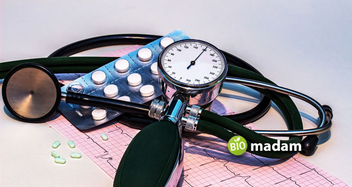
You might have seen doctors using a pump-like device (sphygmomanometer) to monitor your blood pressure. They place the stethoscope beneath the arm wrap of the sphygmomanometer to listen to the heartbeat at the artery. This enables them to find the systolic and diastolic blood pressure when they hear the Korotkoff sounds till the second reading. It is considered the most accurate way of measuring blood pressure despite digital devices.

Abdominal Sounds
Abdominal sounds often explain the changes in GI motility to evaluate diseases and disorders. The doctor will ask you to lie down on your back so they can observe your abdomen. They might also place a pillow under your head and a positioner under your knees. They will then move the stethoscope to different regions of the abdomen to listen to the sounds in the gastrointestinal tract. A critical application of abdominal sounds is understanding recovery after surgery involving anesthesia. The anesthesia causes the bowel movements in the small and large intestine to slow, impacting the patient’s appetite. Continuous monitoring of the abdominal auscultations can help understand the healing process with the changes in the abdominal sounds.
Arteries Auscultation
Arterial auscultation helps understand blood flow through the arteries, primarily in the arms, neck, and abdomen. You may also study the sounds in the arteries in the abdomen. The doctor can identify bruits using an arteries auscultation that appears as a soft sound in the blood when passing through the arteries. Bruits indicate a narrowing of the artery, causing a disturbed flow. The constriction of arteries may occur in the neck, femur, kidneys, ilium, and temporal artery. Furthermore, they also help assess the pulse; a weak pulse may indicate a blocked artery.
Observing Children
Children are not as grown as adults, so doctors use specialized stethoscopes to study children of different ages. Pediatric stethoscopes are slightly smaller than adult stethoscopes, with a scaled-down head to examine the chest better. Similarly, infant stethoscopes are even smaller, with a diameter of no more than 2.6 cm. They are also not made of latex to avoid any stage of allergic reactions. Neonates are the smallest in size, thus needing the smallest stethoscopes. Stethoscopes with a large chest piece may be uncomfortable and fail to provide accurate results. Neonatal stethoscopes help observe neonates and are only 2 cm wide.
Bowel Sounds Observation
Observing bowel sounds is standard practice by healthcare workers when they suspect an issue in your GIT. The movement of food content, fluid, and gas through the intestines produces sounds. The chest piece allows the doctors, nurses, PCTs, CNAs, CRTs, RRTs, etc., to listen to high-pitched irregular bowel sounds. It is critical in diagnosing conditions like paralytic ileus or bowel blockages.
Liver Measurement
While the most common purpose of the stethoscope is to measure the heart and lung sounds, the measurement of the liver size is a more technical function.
The stethoscope can help determine the size of the liver by measuring the vertical length of the liver from the nipple line. The doctor places the index finger in the nipple line towards the belt level with the stethoscope below the right nipple. When your finger is on the liver, you will hear a faint sound in the stethoscope. These two points show the liver, and thus, you can measure the length of the liver. It helps diagnose issues in the liver functioning, as a size larger than 10 cm may indicate health problems.
Fetal Examination
Stethoscopes help find out issues in children and adults and monitor fetal heartbeat. Doctors may use a fetoscope or a Doppler stethoscope for their purpose. They help understand the position of the fetus in the womb. Doppler stethoscopes are extra sensitive to noise to allow parents to hear the heartbeat.

Veterinary Studies
Veterinary stethoscopes are used to study diseases and infections in animals by observing their heart and chest. Typically these stethoscopes help examine mammals like cats and dogs. Vets may also use infant stethoscopes to check the health condition of smaller animals like rodents and lizards.
The Bottom Line
Stethoscopes are critical to medical examination in humans and animals. Doctors use stethoscopes to study heart and lung functioning and record vitals when conducting an initial medical assessment. Stethoscopes may also help study auscultations in other parts of the body like the small intestine, womb, esophagus, trachea, etc. Sometimes, the doctor may use the stethoscope to measure the size of the liver for quick judgment. The uses of stethoscopes are widespread and help understand the patient’s condition, including cardiac and respiratory problems.
FAQs
What was the stethoscope first used for?
In old times, doctors examined the heartbeat and chest movement by placing their ears on the patient’s chest. Rene Laennec, a French doctor, created the first stethoscope using paper to avoid placing his head over a female patient’s chest. He rolled up a piece of paper to listen to her body sounds from a distance.
Why was the stethoscope important?
Opposed to the first-ever stethoscope, advancements in the build and technology allowed doctors to hear and examine more sounds in the body. The stethoscope enables healthcare professionals today to diagnose or prognose any disease by listening to the sounds of the lungs, esophagus, and bowel movements.
What is the most important part of a stethoscope?
The stethoscope comprises three basic parts, namely the chest piece, earpieces, and tube. The chest piece is considered the most important part of the instrument as it enables you to listen to the auscultations in the body. Doctors use stethoscopes with different types of chest pieces to record accurate readings per need. The chest pieces may vary in size and are built to provide the best observation.

Jeannie has achieved her Master’s degree in science and technology and is further pursuing a Ph.D. She desires to provide you the validated knowledge about science, technology, and the environment through writing articles.

