Recently updated on March 5th, 2024 at 07:46 am
The headway in technology helped doctors and other health activists quickly diagnose several infections and diseases. Nowadays, various diagnostic tools are used to detect or narrow down the root causes of ailments, of which X-ray and MRI are the most common.
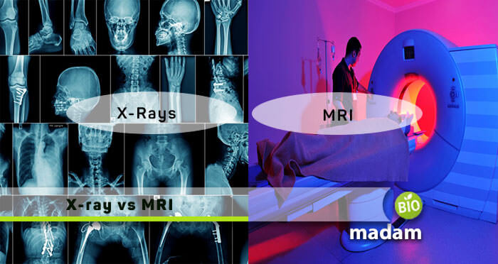
Therefore, we displayed this article to let you grasp the knowledge of both medical imaging technology terms comprehensively.
MRI vs X-Ray – Comparison Table
| Basis of Comparison | X-ray | MRI |
|---|---|---|
| Exposure to Radiations | Harmful ionizing radiations observed | Non-ionizing radiations seen |
| Time for Scan Completion | Within few seconds | Can be 10-15 min. or 2 hours |
| Principle of Application | The attenuated X-rays generates a shadow on the image | Body tissues containing hydrogen atoms assist in emitting radio signals, which the scanner detects |
| Applications | Used for examination of broken bones, identifying diseased tissues, etc. | To estimate soft tissue, such as injury to the spinal cord, brain tumors, etc. |
| Changeable Imaging Plane | Absent | Present |
| Bony Structural Data | Detailed photographs of bone structure on photographic film | Less detailed |
| After Effects | Capable of causing congenital disabilities and diseases, and can also change the DNA | No prominent biological dangers reported |
| Cost | Cheaper | Expensive |
| Soft Tissue Details | Helps observe bones and other dense tissues | Provides details of soft tissue |
| Application Scope | Only detects a couple of body issues | More versatile, and helps examine a variety of medical conditions |
| Image Specification | Differentiate soft tissues and bone density | Differentiate numerous soft tissues types |
What is an X-Ray?
Wilhelm Rontgen was a mechanical engineer and physicist who discovered an X-ray tool in the late 1800s. These electromagnetic radiations penetrate structures differently within the body, hence creating images on a photographic film or a fluorescent screen. We generally call them X-rays Diagnostics that are useful for identifying body abnormalities.
This technology is a painless, non-invasive way of helping to detect problems like broken bones, cancers, dental decay, and foreign bodies. X-rays instantly move through the air and soft tissues of the body. Moreover, they are stopped when denser material, such as a tumor, or bone comes into connection. Diagnostic X-rays are held through a painless operation. So, as a result, positioning the exposed body part between a focused x-ray beam and a film-containing plate. The greater the material density through which it passes, the more rays it absorbs. This way, bone absorbs more x-rays than muscle or fat, and tumors can absorb more x-rays than the tissues around.
What does a Chest X-ray Show?
A chest X-ray uses a small amount of radiation to create images of the structures inside the chest, including the lungs, heart, and surrounding bones and tissues. Here’s what a chest X-ray can show:
- Lungs: A chest X-ray can detect abnormalities in the lungs, including respiratory diseases, such as pneumonia, lung cancer, tuberculosis, or other lung diseases. It can show areas of increased or decreased density, which may indicate fluid, air, or solid masses.
- Heart and major blood vessels: The size and shape of the heart and major blood vessels can be evaluated, which can help detect conditions like an enlarged heart, aneurysms, or other cardiovascular issues.
- Bones: The chest X-ray can reveal fractures, sprains, or abnormalities in the bones of the chest, including the ribs, shoulder blades, and spine.
- Foreign objects: If a patient has accidentally inhaled or swallowed a foreign object, such as a coin or a small toy, a chest X-ray can help locate it.
- Fluid or air accumulation: Chest X-rays can detect the presence of excess fluid or air in the chest cavity, which may indicate conditions like pleural effusion (fluid around the lungs) or pneumothorax (air trapped in the chest cavity).
Chest X-rays are commonly used as an initial screening tool for various respiratory and cardiovascular conditions, as well as for monitoring the progression of certain diseases or the effectiveness of treatments. However, in some cases, additional imaging tests, such as CT scans or MRI, may be required for a more detailed evaluation.
What is an MRI?
Magnetic Resonance Imaging (MRI) is a technology that utilizes a vast magnet and non-ionizing radio waves to observe organs. Doctors in hospitals or technicians in medical laboratories typically apply this technique to scan the soft tissues. Furthermore, it helps us study body structure with no documented long-term side effects. Hence, allowing for broad coverage, ongoing acquisitions, child studies, and adolescents. MRI analysis of the entire body is motivated as per the need for exact determinations of phenotype.
MRI levels have complicated automation in AUs (Arbitrary Units), so the results are intensely affected by homogeneity. The technique, however, wasn’t validated in a whole contiguous body, yet the region has not compared results with those of modalities previously validated.
What does an MRI Show?
An MRI scan, as we know, can provide detailed images of the body’s soft tissues. Here are some of the things an MRI can show:
- Brain and spinal cord: MRI is particularly useful for imaging the brain and spinal cord, as it can reveal structural abnormalities, tumors, injuries, or conditions like multiple sclerosis.
- Joints and musculoskeletal system: MRI can visualize joint structures, cartilage, ligaments, and tendons, making it valuable for diagnosing injuries or conditions affecting the musculoskeletal system.
- Abdomen and pelvis: It can help identify abnormalities in organs like the liver function, kidneys, pancreas, and reproductive organs, as well as detect tumors or other lesions in the abdomen and pelvis.
- Breast: MRI is often used in conjunction with mammography for breast cancer screening and diagnosis, as it can detect small tumors that may not be visible on mammograms.
- Soft tissue: MRI is particularly useful for imaging soft tissues, such as muscles, tendons, and ligaments, making it valuable for diagnosing sports injuries or other soft tissue problems.
- Blood flow: MRI can be used to evaluate blood flow and circulation, which can help diagnose conditions like stroke, heart disease, or vascular problems.
However, it’s important to note that MRI is generally not suitable for patients with certain types of metal implants or devices due to the strong magnetic fields involved.
What is the Difference between an X-Ray and an MRI?
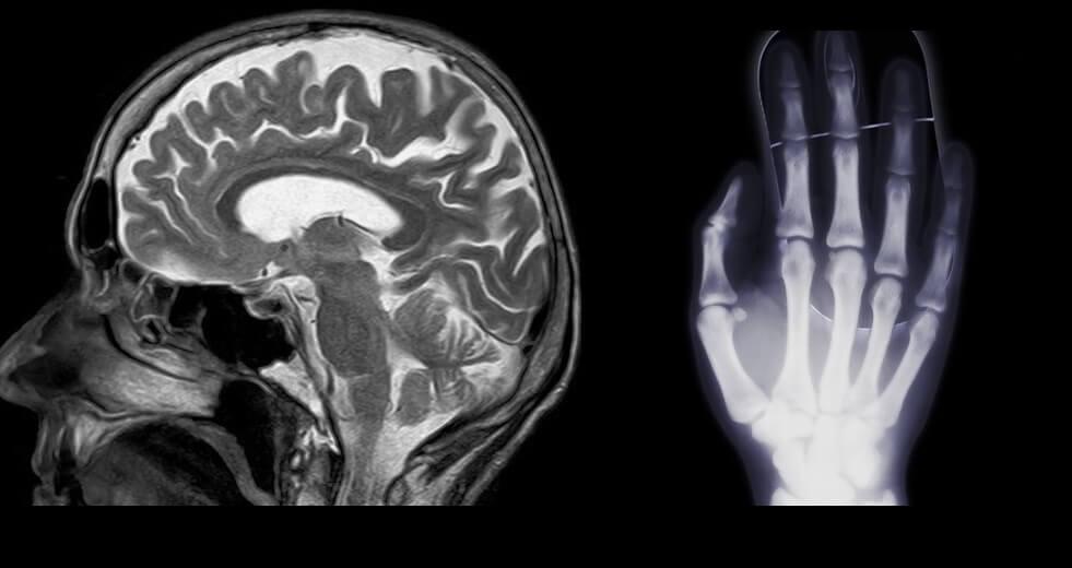
We have enlisted the significant differences in detail below, so let’s begin with its pictorial representation.
Pictorial Variation

Abbreviation
X-Ray
X-ray stands for X-radiation, which is a type of electromagnetic radiation with a wavelength shorter than that of ultraviolet light.
MRI
MRI stands for Magnetic Resonance Imaging, which is a medical imaging technique that uses strong magnetic fields and radio waves to generate detailed images of the body’s internal structures.
Difference in its Application
X-Ray
X-rays are high-frequency beams (wavelengths ranging between 10 and 0.1 nm) of the electromagnetic spectrum, which can easily pass through low-density material (atomic number) but not through high-density substances. So, solid objects such as kidney stones and bones come out very obvious in the X-Ray picture. Click here to check the kidney stone size chart.
MRI
It uses an oscillating magnetic field that is perpendicular to a powerful magnetic theory field. This platform explicitly scans the organ causing issue. Furthermore, the oscillating field in MRI makes magnetization of the hydrogen atoms in a direction perpendicular to the magnetic field of principle. MRI also helps diagnose knee problems, joint pains, and many more, such as:
- Brain issues
- Breast cancer
- Heart diseases
- Wrist dislocation
- Blood vessels, etc.
MRI is a high-speed machine; thus, earplugs or headphones typically support the more bearable sound.
Difference in Techniques
X-Ray
X-rays use radiation to present an image of the internal body structure. Besides, we usually employ them for diagnosing and assessing bone degeneration, fractures, dislocations, infections, cysts, and tumors.
MRI
MRI combines a powerful radio wave magnet (using no radiation) and a computer to create highly detailed images of body structures. The photos appear as cross-sections of the body part being scanned or “slices.” Moreover, MRI scans also assist in treating issues, such as with the bones and joints.
Scanning Duration and Results
X-Ray
X-ray examinations are generally quick, taking only a few minutes for most routine procedures. The actual time spent is usually just a few seconds, with additional time needed for positioning and preparation. Besides, its results are often available more quickly than MRI results, as the interpretation process is generally faster.
- In many cases, the radiologist can provide a preliminary report within a few hours, especially for urgent or emergency situations.
- For routine X-rays, the final interpreted report is typically available within 1-2 days, but it can take longer in some cases, depending on the facility’s workflow and the radiologist’s availability.
MRI
The length of an MRI scan typically ranges from 15 minutes to an hour or more, depending on the area of the body being scanned and the specific type of MRI examination. More detailed or comprehensive scans, such as those of the brain or spine, may take longer, sometimes up to 90 minutes. When getting the results, the time it takes can vary from a few hours to several days, depending on the urgency of the situation and the facility’s workflow.
- In emergency or urgent cases, the radiologist may provide a preliminary report within a few hours.
- For non-urgent cases, the final interpreted report from the radiologist is typically available within 1-3 days, but it can take longer in some cases.
Difference in Hazardous Effects
X-Ray
The X-ray techniques use heavy and dangerous radiations that obviously pose serious health threats. The long exposure of such radiations to bodies can often cause cancers. Furthermore, bodies undergoing X-ray treatments are more prone to damaged soft tissues. The rays emitting from the tool are so heavy that they can even separate the electrons out of the atoms and produce radical ions. Last but not least, these radiations can change a human body’s DNA, thus, altering its specificity.
MRI
On the contrary, MRI is a pretty safe procedure than X-rays. They produce a couple of different cross-sectional images, clicked at the same time. Hence, MRI has a lesser probability of causing complex issues. So far, till now, no biological problem is being reported.
Difference in Usability Limitations
X-Ray
Despite being a well-established and successful imaging technology, X-ray still appears with usability restrictions. It’s especially prohibited to apply to infants keeping in mind the associated risks.
MRI
On the other hand, this technology is restricted to patients with pacemakers or who do not have enough money to afford it. MRI is comparatively an expensive procedure, so experts utilize it less. Otherwise, it has some benefits rather than serious side effects.
Summary
We have differentiated and thoroughly understood the MRI vs X-ray techniques. These two applications come with their beneficial and harmful effects to raise awareness. Still, it’s not up to the patient but the doctor who will tell you which treatment to undergo. But, overall, the imaging technologies, X-ray, MRI, and CT Scan are commonly used to detect different diseases.

Hello, I would like to introduce myself to you! I am Chelsea Rogers, an experienced blog writer for science articles, holding an MPhil degree. My enthusiasm to grab the best knowledge, let it relate to botany, zoology, or any other science branch. Read my articles & let me wait for your words s in the comment section.

