Recently updated on October 4th, 2023 at 09:16 am
Facial bones are an intricate and fascinating part of our anatomy, ranging from the prominent cheekbones that define our face to the delicate bones that protect our eyes and nasal cavity. Facial bones are remarkable body parts that has a crucial role in overall anatomy and physiology. They are essential in establishing our facial structure and supporting the soft tissue that completes our physical appearance. Zygomatic bones are also facial bones consisting of cartilage.
They consist of elastic and hyaline cartilage and are present even in the womb. It then transforms into bone shortly after birth. Zygomatic bones constitute majorly to facial construction issues due to their size and role in joining many facial bones. Hence, it is crucial to acquire knowledge of them. This article will tell you everything about the function of zygomatic bones, structure, and their impact on facial appearance.
What is Zygomatic Bone?
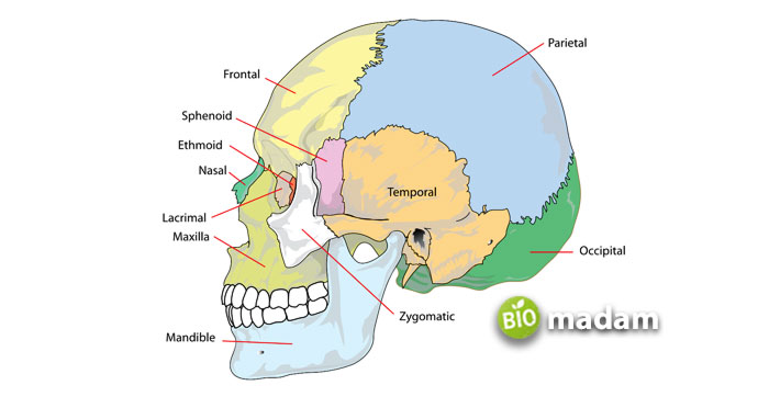
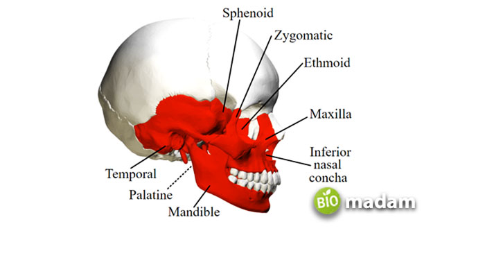
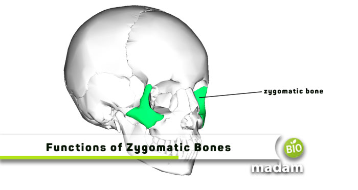
The zygomatic bones are a pair of facial bones that connect the orbit to the lateral skull wall. They are also known as the malar bones and cheekbones. It is one of two irregular bones in the human skull that connect the maxilla to the temporal bone, sphenoid bone and to the frontal bone. They can be found laterally to the maxilla beneath the orbit.
Structure of Zygomatic Bones
The structure of the zygomatic bones is quite intricate, including the zygomatic arch, the temporal process, the orbital process, the maxillary process, and the main body. Near the center of the malar surface is a small opening called the zygomaticofacial foramen. The zygomaticofacial nerve and vessels pass through this region. A slight elevation just below this foramen serves as the muscle’s point of origin, the zygomaticus. Its location is to the side of the maxilla, below the orbit. The temporal, orbital, maxillary, and zygomatic arch processes are part of the bone’s main body.
The broad, flattened extension of the temporal bone, known as the temporal process, articulates with the zygomatic process. The orbital process forms the infraorbital margin and the lateral part of the orbit. The zygomatic arch joins the temporal bone to the body of the zygomatic bone, and the maxillary process articulates with the maxilla bone. The zygomatic bone has a smooth exterior and a cancellous, marrow-filled interior. Small foramina allow blood vessels, including arteries and veins, and nerves to enter the bone and protect it from the environment. The zygomatic bone’s intricate design serves it well in its many roles in the skull and face.

Functions of Zygomatic Bones
Structural Support
The zygomatic bone helps keep the cheeks in place and the general outline of the face intact by forming a bony ridge. The cheeks’ framework comes from the zygomatic bone’s ridges, which give the face its fullness.
Articulation and Muscular Attachment
The zygomatic bone connects with the maxilla, frontal, and temporal bones, among others, through articulation. By joining with it, the lower part of the bone allows the upper jawbone to move and perform functions such as speaking, chewing, drinking, coughing, and breathing that it otherwise could not. These joints stabilize the skull and also control facial expressions.
Protection
The zygomatic bone is an orbital barrier to safeguard the eye. It does double duty in preventing head injuries by dispersing and soaking up any force directed at the face. The pterion is a relatively vulnerable location within the temporal fossa where numerous cranial bones intersect; the zygomatic arch provides some protection. It also shields the internal organs, vascular system, and nervous system.

Aesthetic Appearance
The zygomatic arch often protrudes, leading to a broad look. Zygomatic arches can also cause high cheeks, which is the ideal bone structure for models. A zygomatic reduction procedure can narrow the jawline, and aesthetic specialists can insert an implant or filler to raise the cheekbones.
The Bottom Line
Zygomatic bones are present in facial bones that connect the eye socket to the lateral skill wall. The main functions of zygomatic bones include supporting the facial structure and attachment of muscles. They also protect the eye and improve the aesthetic appearance of your face. Facial trauma or injury may damage zygomatic bones and cause facial swelling.
FAQs
What is the function of the zygomatic process of temporal bone?
The zygomatic process of the temporal bone provides a place for muscle attachment used for chewing and helps strengthen the cheekbone area.
What is the zygomatic bone of the skull?
The zygomatic bone is a diamond-shaped bone lateral to the eye socket at the widest part of the cheek. It plays a role in connecting the frontal bone at the outer edge of the eye socket to the maxilla and sphenoid.
What happens if the zygomatic bone is damaged?
Trauma or injury to the zygomatic bone may not cause any pain for a few days, but it might limit the movement of your mouth. Prolonged damage to the zygomatic bone can lead to pain and sensory damage with displaced cheekbones.
What do zygomatic branches do?
The zygomatic branches present in the face supply part of the orbicularis oculi muscle used to close the eyelid. It makes it possible for the bones to show movement when needed.
What are the 3 components of zygomatic arch?
The three components of the zygomatic arch include the temporal bone, the temporal process of the zygomatic bone, and the zygomaticotemporal suture.
What is the blood supply of the zygomatic bone?
Zygomatic bones in the zygomatic arch get the blood supply from the maxillary artery and its infraorbital branch. Vessels and branches of the arteries in the face nourish the periosteum.
Which bone allows for tears to drain from the eye?
The lacrimal bone located in the medial wall of the orbit provides support to the lacrimal apparatus, which secretes tears. It also acts as a site for orbicularis oculi muscle attachment.
What is the development of zygomatic bone?
The zygomatic bone has evolved from the circumorbital series of bone present in the dermatocranium. These bones develop intramembranous as part of the maxillary prominence.
Is the zygomatic bone strong?
The zygomatic bone is a strong and thick bone with multiple regions of adjacent facial bone articulation vulnerable to external forces. Facial injuries may cause zygomatic-orbital fractures, the most common fractures after mandibular.

Hello, I would like to introduce myself to you! I am Chelsea Rogers, an experienced blog writer for science articles, holding an MPhil degree. My enthusiasm to grab the best knowledge, let it relate to botany, zoology, or any other science branch. Read my articles & let me wait for your words s in the comment section.

