Humans are complex organisms with numerous tissues and organs making up their anatomy and physiology. The skeletal system is one of the most critical elements of the human body. It supports body organs and enables movement along with the muscular system. The skeletal system comprises bones, cartilage, ligaments, and tendons. We often hear about a hyaline fracture in the hyaline cartilage. So, what is it exactly, and what is the function of hyaline cartilage? Keep reading to know it all.
What is Cartilage?
Cartilage is a flexible yet strong connective tissue that protects your bones and joints and helps your bone absorb shocks without damage. It is also present at the end of the bones to reduce friction when you move or use your joints. The cartilage is not as rigid as bones and may receive damage due to trauma or a sports injury. A buildup of cartilage in your joints can cause osteoarthritis.
What is the Hyaline Cartilage?
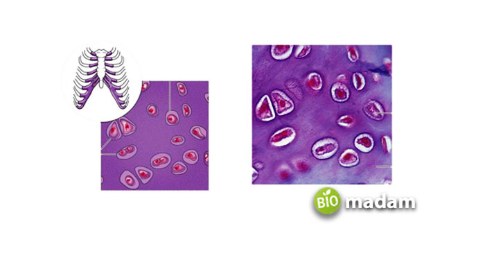
“Hyaline” comes from the Greek word “Hyalos,” which means glassy. This name is given to this cartilage following its shiny appearance. Hyaline Cartilage is the most common in the body and has a smooth, glossy appearance. It looks white and semi-transparent with a tinge of blue shade. It is found around the bones of ball and socket joints or free-moving joints. It is also present in the walls of the respiratory tract, including the nose, bronchi, trachea, and tips of ribs.
Function of Hyaline Cartilage
Smooth Movements
The presence of hyaline cartilage offers smooth movement between joints by reducing friction. It absorbs and withstands pressure that may damage the joints during weight-bearing activities.
Temporary Embryonic Cartilage Model
The hyaline cartilage acts as a temporary cartilage model during embryonic development. These models act as a crucial precursor to appendicular and axial skeleton development.
Shock Absorbance
The hyaline cartilage tissue has a low amount of fiber, allowing it to act as a shock absorber and cushion where joints meet.
Respiratory Support
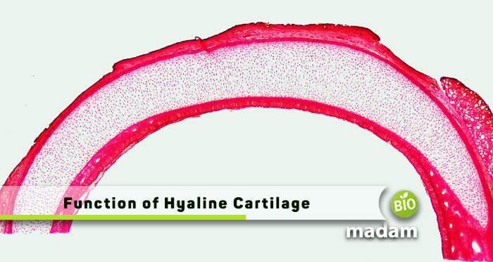
Hyaline cartilage is present in the nose, trachea, and bronchi and supports the respiratory system.

Functions of Hyaline Cartilage in Other Animals
Besides providing respiratory support and acting as a shock absorber in humans, hyaline cartilage is found in many other organisms. The functions of the hyaline cartilage in other organisms include:
- Sharks and rays have a skeleton comprising cartilage as it is less dense than bones and facilitates faster movement in the water.
- Invertebrates like snails and squids have hyaline cartilage that provides structure and support.
- Octopuses have cranial cartilage similar to the hyaline cartilage, one of the only rigid parts of an octopus.
- Gastropods contain the odontophore, which provides feeding support formed from cartilage. It contains a rich amount of myoglobin and some collagen.
Structure of Hyaline Cartilage
The hyaline cartilage is in the extracellular matrix and is a widely distributed connective tissue. It is found in the Laryngeal cartilage, Nasal cartilage, costal cartilage, bronchial cartilage, articular cartilage, epiphyseal cartilage, and tracheal.
It comprises collagens and develops through calcification, the same bone formation process. Cartilage is made of chondroblasts which produce chondrocytes, extracellular matrix, and collagen fibers. The cartilage develops from mesenchymal cells present in the bone marrow. The extracellular matrix is rich in glycosaminoglycans (GAGs), comprising long polysaccharides made of amino sugars.
The hyaline cartilage contains high type II collagen, proteoglycans, and structural glycoproteins. Structural proteins such as aggrecans use link proteins to make covalent bonds with hyaluronic acid. Chondronectin acts as a multi-adhesive glycoprotein that connects to glycosaminoglycans. It also facilitates the binding of chondrocytes to the extracellular matrix.
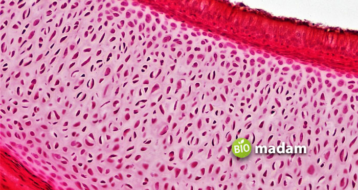
Small amounts of chondrocytes are present in hyaline cartilage mass. They are present in lacunae within the ECM besides type I, IV, V, VI, IX, and XI collagen. Collagen strengthens the fibers to produce cartilage. Every cartilage cell has one nucleus or two nuclei to perform its specified nucleus functions. The membrane surrounding the cartilage is a perichondrium which supplies nutrients to the tissues, as hyaline cartilage does not have blood vessels, like arteries and veins.

Other Types of Cartilages
Besides hyaline cartilage, elastic, and fibrocartilage are also found at the edges of bones to reduce friction.
Elastic Cartilage
Elastic cartilage is also known as yellow cartilage and is quite similar to hyaline cartilage. It gets its yellow color from yellow elastic fibers, which make it flexible. It is found in the auditory tube, outer ear, and epiglottis and gives support and shape to these structures.
Fibrocartilage
Fibrocartilage contains white fibrous tissue and cartilage tissue along with type I and type II collagen. These fibrous proteins make them elastic, flexible, and tough. Fibrocartilage is present in between the pubic bones, the temporomandibular joint, and the disks between vertebrae of the spine. These are responsible to provide protection and support against the shock or stress produced in the body.
The Bottom Line
The human skeletal system comprises bones, cartilage, tendons, and ligaments. Hyaline cartilage has a smooth, semi-transparent, glossy appearance. It is the least strong of all cartilage types. It is present at the end of the bones to reduce friction when you move or use your joints. The main functions of hyaline cartilage are smooth movements, acting as a temporary embryonic cartilage model, providing shock absorbance, and offering respiratory support. They have a high amount of collagen in their structure, along with glycosaminoglycans and proteoglycans.
FAQs
What is an example of hyaline cartilage?
Hyaline cartilage forms the walls of the respiratory tract, including the nose, bronchi, trachea, and tips of ribs. The hyaline cartilage present around the bones of free-moving joints is known as articular cartilage.
What is the property of hyaline cartilage?
The hyaline cartilage acts as a connective tissue. It is the least rigid of the three types of cartilage; however, firmer than other connective tissues like ligaments and tendons, is pliable and provides a definite structure.
What is the difference between hyaline and elastic cartilage?
Hyaline cartilage has a semi-transparent look and is present between joints to avoid friction. On the other hand, elastic cartilage has numerous elastic fibers compared to collagen fibers in hyaline cartilage.
What uses hyaline cartilage for bone formation?
Bony tissues replace hyaline cartilage after endochondral ossification leading to the formation of bones in the skeleton. These bones are known as endochondral bones.
Is hyaline cartilage weak or strong?
Hyaline cartilage is formed by a combination of numerous thin collagen fibers that give it a definite shape. However, it is the weakest of the three cartilage types in the body.
Is hyaline cartilage immovable?
Hyaline cartilage covers the synchondrosis, which are the primary cartilaginous joints. They do not exhibit a lot of movement and are either slightly moveable or immobile.
How does hyaline cartilage grow?
Hyaline cartilage grows in length due to the secretion of the new matrix within the cartilage. These occur in interstitial growth. Alternatively, appositional growth involves the secretion of the matrix along existing surfaces, which pushes the cartilage to expand in width.

Hello, I would like to introduce myself to you! I am Chelsea Rogers, an experienced blog writer for science articles, holding an MPhil degree. My enthusiasm to grab the best knowledge, let it relate to botany, zoology, or any other science branch. Read my articles & let me wait for your words s in the comment section.

