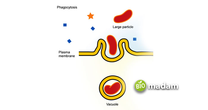
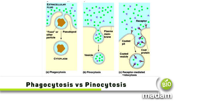
Phagocytosis and pinocytosis are the types of endocytosis, a cellular process that absorbs extracellular material. Hence, it typically helps a cell’s intracellular spaces attain beneficial material from outside. Both of its types are active processes, always requiring energy (Adenosine Triphosphate) to uptake materials.
Phagocytosis represents the eating of a cell, whereas pinocytosis indicates cell drinking. The point that differentiates these two terms is their form of ingestion. The former engulfs large solid organisms, for example, amoeboids, bacteria, protozoa, and other parasites. On the other hand, the latter forms buds to create invaginations and drinks liquid particles. Let’s start by discussing a comparison chart below.
Comparison Table
| Literal Meaning | Cell eating | Cell drinking |
| Engulfing Method | Through false feet (Pseudopodia) | Through invagination |
| Specificity | Substrate specific | Substrate non-specific |
| Type of Material Intake | Solid | Liquid/Fluid |
| Vesicles Formation | Phagosomes | Pinosomes |
| Role of Lysosomes | Yes | No |
| Examples of Engulfed Particles | Microorganisms, Foreign particles, dust, etc. | Minerals, ions, sugar, amino acids, etc. |
| Nature of Process | Triggered | Constitutive |
| Presence/Absence of Exocytosis | Present | Absent |
We’ve elaborated the two terms individually, in detail, so there is no confusion left. Hence, continue reading the article below to know more about phagocytosis vs pinocytosis.
What is Phagocytosis
Elie Metchnikoff first observed the phenomenon of phagocytosis and derived the term from a Greek word, phagein, meaning “to eat.” It helps a cell swallow solid particles in the plasma membrane and ends in forming vesicles called phagosomes. The enzymes of phagosomes digest these ingested particles and break them into simpler units before absorption.
Some examples of phagocytosis are cell debris, foreign antigens, microorganisms, etc. A vesicle typically goes around 1-2 μm in size to enter a cell membrane.
Primary Phagocytosis Function
This mechanism proceeds through special cells called phagocytic cells. Phagocytosis holds significant importance as it functions in innate immunity. Innate is naturally obtained immunity, unlike adaptive immunity which is further subdivided into active and passive immunity. Besides, phagocytosis is the first-line defense against any infection or foreign particle and is the first-line defense against any infection or foreign particle. Secondly, it plays a crucial role in bringing adaptive immune response to a body. Phagocytosis consists of several cells, particularly immune cells, engulfing solid particles, such as neutrophils, monocytes, eosinophils, etc. Let’s briefly understand the functioning of these cells in phagocytosis.
- Monocytes – these phagocytic cells are present in the blood and usually circulate in the blood and spleen. Monocytes make up about 5% to 10% of white blood cells. Furthermore, they phagocytose dead cells and foreign bodies, including pathogens, in response to nitric oxide production. Thus, helping a body develop different types of immunity.
- Eosinophils – are motile cells playing against multicellular parasitic organisms, such as those not easily phagocytosed. Eosinophils form clusters around invading worms that have damaged membranes by protein activity released from eosinophilic granules.
- Neutrophils – are the most abundant WBCs, constituting about 50% to 70% of circulating leukocytes. Neutrophils are also motile phagocytic cells, and Metchnikoff called them the archetypal phagocyte. These immunity cells use reactive oxygen species to phagocytose and also kill harmful pathogens.
- Dendritic Cells – function to phagocytose deadly parasites, bacteria, cell debris, and even intact cells very efficiently.
Phagocytosis Steps
Below are the steps following phagocytosis
- Activation of the Phagocyte
- Chemotaxis of Phagocytes
- Attachment of the Phagocyte to the Extracellular Particles (Microbes or Cells)
- Ingestion of the Microbes or Cells by the Phagocyte
- Destruction of the Microbes or Cells
- Elimination
Activation of the Phagocyte

Inflammatory mediators are active phagocytic cells that are either a whole-cell, e.g., bacteria or virus, or any component of the cell such as bacterial proteins, capsules, prostaglandins, or complement proteins. The phagocytic cell then enters the tissue from the capillaries to reach the infection site. These cells produce Pattern Recognition Receptors (PRRs), involved in analyzing the pathogen and making a bond on the pathogen’s surface
Chemotaxis of Phagocytes
This reaction helps determine the direction of cells’ movement in response to chemical stimuli. Furthermore, it directs the phagocytic cells to the site of infection or the area where a high concentration of foreign particles is present. Different chemotactic molecules, called attractants, are bacterial products, cell and tissue debris, and components of the inflammatory exudate, such as peptides derived from the complement.
Attachment of the Phagocyte to the Extracellular Particles
This step involves receptors present on the phagocytic cell membrane and necessary for the ingested molecule. Different receptors for phagocytosis are used, depending on the cell type. But still, a few are primarily used, such as opsonin receptors, scavenger receptors, toll-like receptors, and antibodies.
Ingestion of Microbes or Cells by the Phagocyte
Soon after the attachment, the cell membrane starts surrounding foreign molecules to trigger ingestion. It happens by the plasma membrane’s infoldings, resulting in a vacuole called the phagosome. The ingestion depends upon the molecule size as the smaller ones (e.g., bacteria) are ingested more instantaneously while the large molecules are phagocytosed more over time.
Destruction/Elimination
In the end, hydrolytic enzymes (phagocytic enzymes) digest the ingested molecules. The cell lysis kills bacteria and then digests them with the help of lysosomal enzymes.
What is Pinocytosis

The word Pinocytosis comes from a Greek Language, Pinos, meaning “to drink.” Warren H. Lewis, in 1931, discovered the process, which is primarily the ingestion of small liquid particles. Thus, it is an endocytosis type where fluid particles from extracellular spaces enter a cell through the pores opened on its membrane. Pinocytosis generates vesicles due to the plasma membrane invagination and is called Pinosomes. It is also referred to as Fluid Endocytosis or Bulk-Phase Endocytosis.
Types of Pinocytosis
This process can be of different types, based on the category.
Based on Molecular Size
Let’s first discuss pinocytosis depending on its molecular size. These are:
- Micropinocytosis
- Macropinocytosis
Micropinocytosis
As the name indicates, this type of pinocytosis takes in smaller molecules. The vesicles formed are about 0.1μm in length, which leaves tiny depressions on the cell surface after entering. Micropinocytosis plays a vital role in transferring molecules across capillary walls, for example, Caveolin-Mediated Pinocytosis.
Macropinocytosis
On the contrary, the name indicates this pinocytosis type intakes larger molecules inside the cell. The vacuoles formed after the foreign particles invade a cell are usually of size 1-2μm. Macropinocytosis is used in migrating cells as it helps the plasma membrane uptake with their direction of motion.
Based on Receptors
Special receptors help continue the process of pinocytosis; hence there are different types of it, depending on receptors. These are:
- Clathrin-Mediated Pinocytosis
- Caveolin-Mediated Pinocytosis
- Clathrin- and Caveolin- Independent Pinocytosis
Primary Pinocytosis Function
It is a non-specific constitutive process, that occurs all the time. Pinocytosis actively helps to intake the surrounding liquid material required for cell survival, e.g., hormones, enzymes, ions, sugars, etc. It usually occurs in almost all body cells, particularly in secreting cells and cell lining of the blood capillaries; besides, in complex living organisms, pinocytosis functions to transfer dissolved fats, proteins, and vitamins in bulk. As the kidneys has a crucial role in eliminating waste through urine, this process plays its part by excreting that waste from the cells.
Pinocytosis Process
Below are the detailed steps following pinocytosis:
Initiation
The process begins when external molecules bond strongly with the cell membrane’s receptor cells, sending clear signals to form inner folding.
Formation of Inner Pockets/Invagination
The cell’s membrane then forms tiny pocket structures or inner folding to absorb extracellular fluid particles. These pockets are open from the ends, but they start closing soon after the liquid enters a cell.
Formation of Vesicles
Once fully engulfed, the animal cell membrane throws the particles inside the cell in tiny packets called vesicles. Besides, these are unique organelles circling the extracellular fluid material. These structures help the external particle move quickly within the cytoplasm of a cell or stay connected to the endosome.
When a vesicle is freely floating in the cell, it reaches the other end to acquire exocytosis. This step is vital to keep up the original size of a cell. In the second case, the fusion of vesicles with endosomes aid in hydrolyzing the particles to tiny molecules for further use.
How do Phagocytosis and Pinocytosis Differ?
Phagocytosis and pinocytosis are two distinct forms of endocytosis, but they differ in the type of materials they ingest and the mechanisms involved. Phagocytosis involves the ingestion of large solid particles such as bacteria, dead cells, or debris by specialized cells like macrophages and neutrophils. The cell membrane extends outward, forming a pocket around the target material, and then pinches off, engulfing the material within a membrane-bound vesicle called a phagosome. In contrast, pinocytosis is the ingestion of small molecules, fluids, and dissolved substances from the extracellular environment. It involves the formation of small, fluid-filled vesicles called pinocytic vesicles that bud inward from the cell membrane, capturing a portion of the extracellular fluid and its contents. Unlike phagocytosis, pinocytosis does not involve the ingestion of large solid particles but rather the uptake of soluble materials and small particles through the formation of these pinocytic vesicles.
Summary
So, there should now be no confusing when understanding questions like, “What is the difference between phagocytosis and pinocytosis.” Both phagocytosis and pinocytosis are essential processes that enhance the particles’ ingestion inside a cell. The only difference in variating them is the nature of the material chosen. Phagocytosis engulfs solid particles, mostly helping in a human’s immunity system. On the other hand, Pinocytosis exists in all body parts to form invaginations for liquid particles.

Hello, I would like to introduce myself to you! I am Chelsea Rogers, an experienced blog writer for science articles, holding an MPhil degree. My enthusiasm to grab the best knowledge, let it relate to botany, zoology, or any other science branch. Read my articles & let me wait for your words s in the comment section.

