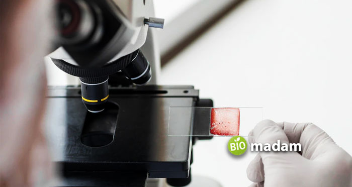
Compound and electron microscopes are widely used in all branches of biology mainly in microbiology and chemistry research labs and schools.
They provide ease to the students and researchers to study micro samples. A light microscope is more common in homes and schools, whereas you may find electron microscopes in advanced research centers only. It is mainly because of the electron microscope size and cost. You can observe cells of different samples, like blood and plasma under a light microscope. However, electron microscopes give a more detailed specimen observation.
Let’s tell you the differences between a light microscope and an electron microscope in detail.
Comparison Table
| Features | Compound Microscope | Electron Microscope |
| Objective | Outer surface of cells | 3D detailed view of specimen |
| Invention | 16th century | 1931 |
| Magnification | 500x – 2000x | 100,000x – 500,000x |
| Resolution | 0.25µm to 0.3µm | 0.001µm |
| Illumination | Halogen, fluorescent, LED | Electron beam |
| Wavelength | 400nm- 700nm | 1nm |
| Lens | Glass lenses | Electromagnetic |
| Specimen Preparation | Few minutes or hours | Days |
| Samples | Alive and dead | Dead and dry |
| Object Thickness | 5µm or more | 0.1µm or less |
| Staining | Stains | Heavy metal |
| Image Color | Colored | Black and white |
| Image Observation | Eyepiece or LCD screen | Photographic plate |
| Image Formation | Light absorption | Electron scattering |
| Vacuum | Absent | Present |
| Power | Standard | 50,000 volts or more |
| Cooling System | Absent | Present |
| Filament | Absent | Tungsten |
| Risk of Radiation Leakage | No | Yes |
| Cost | Low | High |
What is a Compound Microscope?
A compound microscope is a medium-sized microscope that you can easily place on a rack or a table. It takes minimal space. Light microscopes are more common in schools for teaching purposes. Many people also buy them to teach their children about microscopes at home.
A compound or light microscope uses light to illuminate the specimen. The sample is placed on the stage and illuminated by the light source. Light microscopes typically have a halogen, fluorescence, or LED light. Compound microscopes produce magnified images through eyepiece and objective lens combined. The eyepieces generally are 5x and 10x, and sometimes 20x. Their magnification in biology is of high importance. Some microscopes also have an LCD to observe the specimens.
These microscopes examine human skin tissues, cells, bacteria, and viruses. Their magnification and resolution are quite less compared to electron microscopes. However, they can observe live cells of all sizes, which an electron microscope cannot.
Types of Compound Microscopes
Compound microscopes are differentiated into various types of light used for illumination. They are used to produce specific quality images. The most common types of compound microscopes are:
- Darkfield microscopes
- Fluorescence microscopes
- Phase contrast microscopes
- Confocal microscopes
- Polarized microscopes

What is an Electron Microscope?
As opposed to compound microscopes, electron microscopes are large-sized devices and are not found in all laboratories. They use electron beams to illuminate and magnify the object. Knoll and Ruska developed the electron microscope in Germany in 1932, which was first used in 1940.
Researchers use electron microscopes to observe subcellular structures that you may not see through a compound microscope. Electron microscopes magnify images up to 500,000 times. The sample must be dry and ultra-thin. It uses an internal vacuum and high-voltage electricity to run a current in the machine. The only flaw (as some may call it) is that the electron microscope cannot observe live cells. You can observe viruses on surfaces though.
Types of Electron Microscopes
Electron microscopes have three types according to electron bombardment and reflection techniques. They are used for different purposes depending on the sample and the required observation. The three major types of electron microscopes are:
- Scanning electron microscope (SEM)
- Transmission electron microscope (TEM)
- Reflection electron microscope (REM)
Differences between Compound and Light Microscope
Objective
Compound Microscope
Compound microscopes help observe the outer surface of cells at the cellular and tissue levels like epithelial and connective tissues.
Electron Microscope
Whereas electron microscopes aid in studying the gross internal 3D structure of cells.
Invention
Compound Microscope
Compound microscopes were first manufactured in the 16th century by Zacharius Jansen and Hans.
Electron Microscope
German engineer Max Knoll and Physicist Ernst Ruska developed the electron microscope in 1931.
Magnification
Compound Microscope
Compound microscopes have magnifications ranging between 500x to 2000x. You can calculate microscope magnification with this technique.
Electron Microscope
Conversely, electron microscopes have a high magnification of 100,000 to 500,000x.
Resolution
Compound Microscope
Compound microscopes have a low resolution ranging between 0.25µm to 0.3µm.
Electron Microscope
On the other hand, electron microscopes have 0.001µm resolution, which is 250 times higher than compound microscopes.
Illumination
Compound Microscope
Compound microscopes work with a light source to illuminate the object. It is a halogen, fluorescence, or LED source.
Electron Microscope
In contrast, an electron microscope uses a beam of electrons to illuminate the sample.
Wavelength
Compound Microscope
Compound microscopes use the light of wavelengths between 400nm – 700nm.
Electron Microscope
Unlike, electron microscopes use a low wavelength produced by electrons of around 1nm.

Lenses
Compound Microscope
Compound microscopes have glass lenses – condenser, eyepiece, and objective lenses.
Electron Microscope
Electron microscopes do not have glass lenses. Instead, the focusing lenses are electromagnetic.
Specimen Preparation
Compound Microscope
It takes only a few minutes or hours to prepare a slide for compound microscope observation.
Electron Microscope
On the other hand, it may take days to prepare a specimen for an electron microscope.
Samples
Compound Microscope
You can observe all kinds of live and dead samples in a compound microscope.
Electron Microscope
At the same time, an electron microscope only studies dead and dry samples in a thin-sheet-like form.
Object Thickness
Compound Microscope
The objects observed under a microscope are usually 5µm thick or more. They are suitable for gram-positive and gram-negative bacteria slides.
Electron Microscope
In comparison, objects for electron microscope should be 0.1 µm or less in thickness.
Staining
Compound Microscope
Specimens studied under a compound microscope are typically stained using colored dyes.
Electron Microscope
Alternatively, electron microscopes require specimen coating with heavy metal to reflect electrons.
Image Color
Compound Microscope
The images produced by the compound microscope are the same color as the specimen or the stain. You can observe colored images through a light microscope.
Electron Microscope
While electron microscopes give a black-and-white image due to electron bombardment.
Image Observation
Compound Microscope
The magnified image of a compound microscope is studied through the eyepiece lens or a digital LCD screen.
Electron Microscope
Though the images of an electron microscope appear on a photographic plate or Zinc Sul pate Fluorescent Screen. They can also observe archaea and eubacteria in depth.
Image Formation
Compound Microscope
The image formation in a compound microscope depends on light absorption from varying zones.
Electron Microscope
However, the images by an electron microscope depend on the electron scattering.
Vacuum
Compound Microscope
A compound microscope operates through light and does not need a vacuum.
Electron Microscope
On the opposite, the electron microscope requires a vacuum to produce images of the specimen through an electron beam.
Power
Compound Microscope
A component microscope can easily function anywhere without the need for high voltage.
Electron Microscope
But, electron microscopes need high voltages of up to 50,000 volts or more.
Cooling System

Compound Microscope
Due to less heat emission from the light source, compound microscopes do not require a cooling system.
Electron Microscope
On the contrary, electron microscopes require high voltage and produce a large amount of heat. A cooling system keeps the instrument running smoothly.
Filament
Compound Microscope
No filament is present in compound microscopes.
Electron Microscope
Yet the filament is a vital part of the electron microscope to produce the electron beam.
Radiation Leakage
Compound Microscope
As compound microscopes use light radiation, there is no risk of radiation leakage.
Electron Microscope
However, radiation leakage risk exists when using an electron microscope.
Cost
Compound Microscope
Compound microscopes come in multiple price ranges, and you can buy microscopes for under $100. The maintenance cost is also low.
Electron Microscope
Conversely, the electron microscope is an expensive instrument with high maintenance costs.
The Bottom Line
Microscopes are an important part of studying specimens in biology and research labs. Compound microscopes are more suitable microscopes for students and learners, while electron microscopes are more widely used in research and development. Light microscopes provide a view of the outer surface of cells and tissues. On the other hand, electron microscopes study the 3D structure of the specimens in detail. They have high magnification and are more costly. The above-mentioned list of differences between compound and electron microscopes will help you to choose the right one for your needs.

The Biomadam Content Team develops and manages educational and informational articles published on Biomadam. Content is prepared using standard reference materials and follows internal editorial guidelines to ensure clarity and consistency.
