Recently updated on March 5th, 2024 at 11:13 am
Every human being is blessed with different regulatory systems, including nervous system, endocrine system, circulatory system, etc. The complex nervous system comprises the brain, sensory organs, spinal cords, and neurons. These function to connect different nervous systems, including the central and the peripheral nervous system, with other parts of the body, and also transmit signals. The neurons particularly have:
- Dendron (dendrites)
- Axon
- Cell body
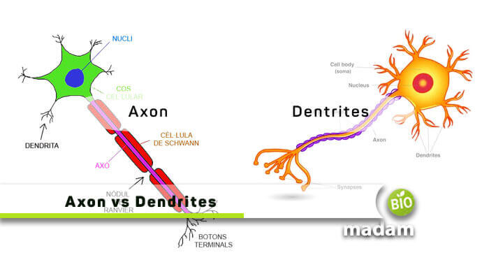
Dendrons and axons are involved in the conduction of nerve impulses through the nerve cell. Moreover, both are unique cytoplasmic projections of neurons. If a dendrite brings information to the cell body from the other neurons, the other (axon) takes information from the body. There are a lot more differences that we will look into in this article. First, check out the comparison table listed below!
Comparison Table
| Basis of Comparison | Axon | Dendrites |
|---|---|---|
| Type of Signal | Signal Transmitter | Signal Receptor |
| Length | Longer | Comparatively Shorter |
| Origin | From an Axon Hillock | Directly from Nerve Cells |
| Number of Cells | A single axon in a neuron | Multiple dendrites in a neuron |
| Surface Nature | Smooth | Rough (Dendritic Spines) |
| Myelin Presence | Present | Absent |
| Branched At | Distal End | Everywhere |
| Neurotransmitter Containing Vesicles | Present | Absent |
What is an Axon
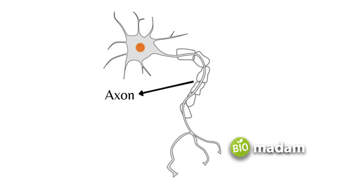
An individual, most extended nerve cell projection is called an axon. It mainly functions to carry impulses away from the cell. Axons are originated from specialized regions further called axonal hillocks. In addition, they may sometimes arise from the dendrites. The axon’s proximal part adjacent to the axon hillock is the initial segment that starts right after the axon hillock.

These cell types are further branched at the distal end, which is usually swollen. There are abundant proteins present in axons, primarily containing the microtubules and microfilaments. As a result, the special axonal microtubules are aligned with the long axis of the axon. They have a uniform polarity that is distal to the soma. Microtubules also play a critical role in the transport of metabolites and organelles in the axon. Besides, they have a highly phosphorylated cytoskeleton.
What Does the Axon Do?
Axons send their signals through a unique axonal transport procedure, working certainly for the growth and survival of a living body. It is the active process by which proteins and other substances get synthesized in the neurosome and are also transported further. Axonal transport is of the following types:
- Fast Axonal Transport (up to 400 mm/day): It is employed during development to grow axons and dendrite. It is elongated by adding new material to its tips.
- Slow Axonal Transport (5 mm/day): It is also known as axoplasmic flow.
What are Dendrites
On the other hand, dendrites are short extensions that carry nerve impulses towards a cell body from the synapsis. A single cell body gives rise to numerous dendrites. Hence these are highly branched structures. A cluster of dendron occupies a large surface area, around 300,000 μm3 of the neuron (motor neuron), and provide 370,000μm3 for synaptic input. Like an axon, dendrites also have several organelles, such as microtubules, microfilaments, functioning of ribosomes, etc. These enable them to alter protein density in response to changes in the frequency of neuronal inputs and help in the regular activity of neurons.

If we consider the different organelles of a dendritic cell, its free ribosomes are present throughout the cytoplasm. Moreover, the smooth endoplasmic reticulum situated helps in the regulation of cytoplasmic calcium. Besides, it has a cytoskeleton that is poorly phosphorylated, unlike an axon. Some other unique dendritic organelles are dendritic spines and dendritic swellings.
What Do Dendrites Do?
They serve a crucial function in receiving and processing incoming signals from other neurons or sensory receptors. Here are the main roles of dendrites:
- Dendrites are covered with tiny protrusions called dendritic spines, which act as the receiving sites for synaptic inputs from other neurons. When a neurotransmitter is released from the presynaptic neuron, it binds to receptors on the dendritic spines, initiating a postsynaptic response.
- Dendrites integrate the various synaptic inputs received from different presynaptic neurons. The spatial and temporal summation of these inputs determines whether the neuron will generate an action potential (nerve impulse) or not.
- These extensions actively process and modulate incoming information through various mechanisms, such as dendritic spines, ion channels, and neurotransmitter receptors.
- Dendritic spines can undergo structural changes in response to patterns of synaptic activity, which is believed to be the cellular basis for learning and memory formation. This process, known as synaptic plasticity, involves the strengthening or weakening of synaptic connections.
Axon vs Dendrite – Major Differences
We will further continue with the apparent differences between the two terms.
Definition
Axon
Axons are derived from the Greek language, also called nerve fibers, as they appear elongated and slender with uniform thickness and smooth surface throughout the length.
Dendrites
The dendritic name comes from the Greek word “Dendron,” meaning “tree.” These are the receptive surface of the neuron and are also termed as the output devices. In the mature nervous system, the dendritic branching of a neuron is changeable, either growing or retracting.
The Direction of Electrochemical Impulses
Axon
They are protoplasmic projections of nerve cells arising from the discharge end of the nerve. Secondly, axons carry electrochemical impulses away from the cell body of neurons.
Dendrites
These are also membranous tree-like protoplasmic projections arising from the body of a neuron that operates through signaling from other neurons (electrochemical impulses).
Presynaptic/Postsynaptic Terminals
Axon
These neuron types are presynaptic in the branched function at the distal end, often producing hundreds or thousands of presynaptic terminals.
Dendrites
The impulses they receive transports them inwards and towards the soma or cell body. They are shorter and postsynaptic as compared to axons.
Length
Axon
A neuron typically contains a single axon that varies in length depending on its type. As compared to the dendrites, these may be several meters long, above 50mm. Axons in neurons are long to carry information throughout the body. The axon of the sciatic nerve is the most extended in the human body, which runs from the spinal cord and reaches the big toe of each foot.
Dendrites
The size and complexity of dendritic arbors increase during development, which is almost 5-7 per neuron. Furthermore, the length of a dendrite goes to 2μm, most of the time.
The Shape of the Neurons
Axon
The axonal shape doesn’t change, where its radius remains constant.
Dendrites
In contrast, such nerve cells usually have tube-like bodies that are typically tapered.
Nature of Biosynthesis
Axon
These structures are mainly rich in smooth ER, however, rough ER is also present in low quantities. Thus metabolic activity (biosynthesis) in axons is limited. But at the same time, they are enriched with low molecule weight microtubules associated proteins.
Dendrites
Also called cell bodies, dendrites have high molecular weight microtubule-associated proteins designated MAP2. Dendrites also possess organelles such as plasma membrane, mitochondria, vacuoles, neurofilaments, microtubules, ER, and ribosomes. These organelles are the sites of protein synthesis that help in memory formation.
Abundant Proteins
Axon
The most abundant proteins in the axon are the proteins forming microtubules, neurofilaments, and actin filaments (microfilaments). Microtubules are a dominant feature of all axons.
Dendrites
On the contrary, there are many dendrites per neuron and arise from the receiving end of the neuron. They receive the impulses via synapses and contain specialized proteins that accept processes and additionally transfer these to the cell body.
Myelinated/Non-Myelinated
Axon
Some axons have an insulating layer called myelin sheath made of protein and fatty substances in the vertebrates, a subphylum of chordates. It protects the axons from electrical impulses. In addition, the myelin sheath speeds up the nerve transmission. In large axons, transmit impulses transmit at speeds up to 90 meters (300 feet) per second.
Dendrites
Unlike axons, these neurons do not contain any myelinated sheath for insulation.
Synaptic Knob
Axon
The axon terminal forms an enlarged synaptic knob that links one neuron part (usually its axon) and the dendrites, cell body, or axon of a second neuron. A neurotransmitter is present in the synaptic knob, such as acetylcholine, norepinephrine, and dopamine.
Dendrites
On the contrary, there are no synaptic knots on the tip of dendrites.
Neurofibrils & Nissl’s Granules
Axon
Axons contain neurofibrils all over, but they lack Nissl’s granules.
Dendrites
They contain both neurofibrils and Nissl’s granules. The complexity of the dendrites reflects the number of connections that a neuron receives.
Conclusion
Axon and dendrites are like any other essential part of a human body that have similarities and differences too. Above all, both are the nerve cell’s projections whose primary function is to transmit nerve impulses. These branched structures are also common in consisting of several neurofibrils. Despite everything, the primary difference between an axon and a dendrite will still remain in its direction of transmitting an impulse.

Hello, I would like to introduce myself to you! I am Chelsea Rogers, an experienced blog writer for science articles, holding an MPhil degree. My enthusiasm to grab the best knowledge, let it relate to botany, zoology, or any other science branch. Read my articles & let me wait for your words s in the comment section.

