Before we head towards our main topic, the classification of epithelial tissue, let’s first understand the term.
Epithelial Tissues: The thin tissues lining the interior of hollow organs and covering exposed body surfaces such as skin, airways, digestive tracts, urinary, and reproductive systems. These issues are one of the four major tissue types in animals. Epithelial tissue consists of epithelial cells arranged in a continuous sheet, in either single or multiple layers underlined by the basement membrane. The basement membrane is an extracellular matrix, that functions to support epithelial tissue structurally. It further consists of two layers, named the basal lamina and the reticular lamina. The basal lamina is the mixture of proteins such as glycoproteins, laminin, collagen, and proteoglycans secreted by the epithelial tissue on which they sit and help the epithelial cells hold one side of connective tissue. These structures typically are made up of fibrous or globular proteins.
No blood vessels cross the basement membrane to enter the tissue because they are completely a-vascular. Nutrients must come by different types of diffusion or absorption from underlying tissues or the surface inside the tissue.
Epithelial cell membranes have the following three regions, distinct in function & structure that additionally provide membrane polarity.
- Apical
- Lateral
- Basal
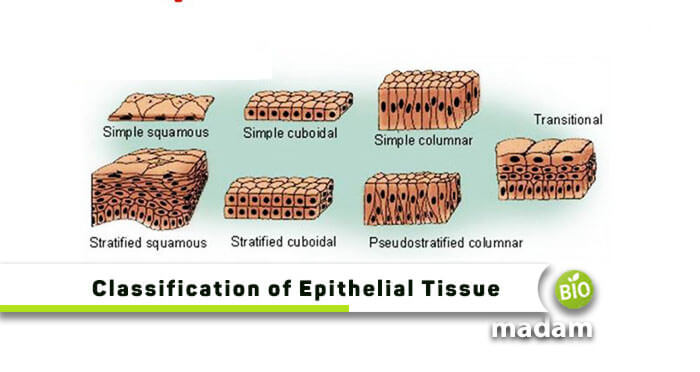
Classification of Epithelial Tissues
These tissues are classified on different criteria, including several cell layers, cell shape, cell secretion, and of course, simplicity or complexity of tissues. We can differentiate epithelial tissues as:
- Simple Epithelial Tissues
- Compound Epithelial Tissues
- Glandular Epithelial Tissues
Simple Epithelial Tissues
These tissues have only one layer of cells attached to the basement membrane. They help in the body’s osmosis and diffusion to further facilitate filtration, absorption, and secretion processes. Simple epithelial tissues have further sub-classification as the following:
- Simple Squamous Epithelium
- Simple Cuboidal Epithelium
- Simple Columnar Epithelium
Simple Squamous Epithelium
The simple squamous epithelium consists of a single layer of flat cells and thin scales. Epithelial tissue lines vessels of the lymphatic and cardiovascular system and comprises a single layer of squamous cells called the endothelium. Its function is to secrete a lubricating substance, allowing diffusion and filtration. This squamous epithelium is present wherever the rapid passage of chemical compounds is observed because of its thinness.
These cells are present in blood and lymphatic vessels (lining of capillaries), air sacs of lungs (alveoli), the lining of the heart, glomerular (Bowman’s capsule) of kidneys, and the inner surface of the tympanic membrane (eardrum).
Simple Cuboidal Epithelium

Simple cuboidal epithelium is a single layer of cube-shaped cells and has a nucleus of box-like cells that appears round and is generally located near the center of the cell. Furthermore, it allows the excretion, secretion, or absorption of molecules. Simple cuboidal epithelia prevail in the lining of the kidney tubules, and ducts of small glands and cover the surface of the ovary. Besides, it also lines a capsule’s anterior surface and eye’s lens, forming pigmented epithelium at the posterior surface of the retina of the eyes and the surface of the ovary.
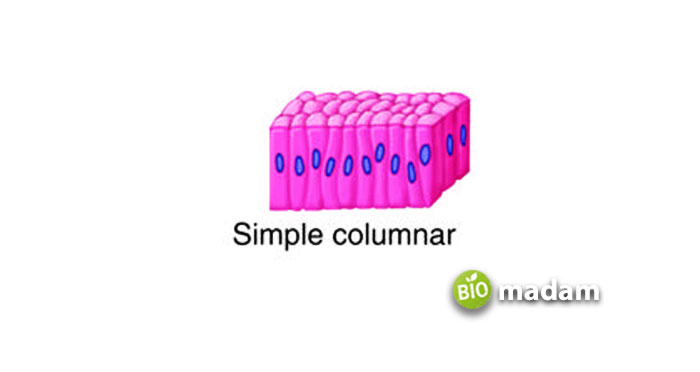
Simple Columnar Epithelium
It is another simple epithelial tissue consisting of a single layer of closely packed elongated cells located in the cells’ basal end. These tissues can be further classified based on the presence of motile cilia, and ciliated or non-ciliated columnar epithelium. Ciliated tissues, including the respiratory system (bronchi), help to remove particulate matter, uterine tubes, and uterus. Non-ciliated is present in the digestive system (lining the stomach and the small intestine, with microvilli and parts of the female reproductive tract. The columnar epithelium is active in the absorption and secretion (secrete mucous and enzymes) of molecules and provides a barrier against the non-specific movement of luminal substances.
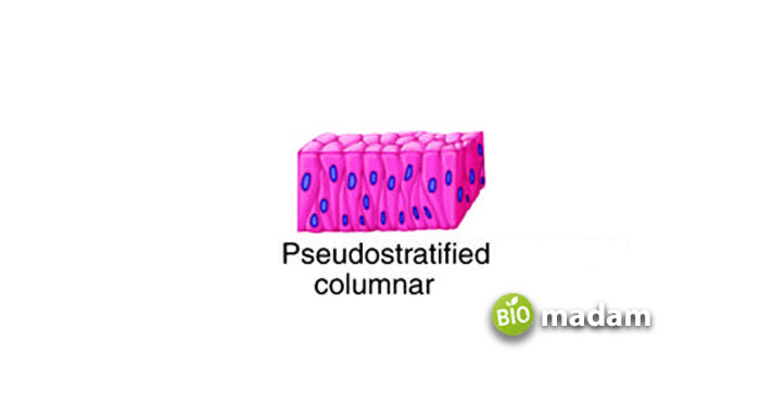
Pseudostratified Epithelium

It is a sub-type of columnar epithelium that contains a tuft of cilia tops in each cell. Pseudostratified columnar epithelia are tissues coming from a single layer of cells made from multiple layers (stratified). In addition, nuclei of neighboring cells appear at different levels rather than clustered in the basal end leading to the illusion of being stratified, and each cell being in contact with the basement membrane. The pseudostratified columnar epithelium is found in the respiratory tract (nasal cavity, trachea, and bronchi). When combined with stereocilia, these are found in epididymis, a highly coiled genital duct of males, larger ducts of many glands, and a part of the male urethra.
Stratified/Compound Epithelial Tissues
Another sub-classification of epithelial tissues is the stratified or compound tissues, having more than one cell layer. These cells are comparatively thicker than the simple ones with the absence of a basement membrane. In contrast, the basal layer is the only one in contact with the basal lamina. The stratified epithelium has a mechanical or protective role against physical and chemical wear and tear. Such tissues have the following sub-types, based on their shapes and cell layer:
- Stratified Squamous Epithelium
- Stratified Cuboidal Epithelium
- Stratified Columnar Epithelium
- Stratified Transitional Epithelium
Stratified Squamous Epithelium
It has two or more squamous cells at the superficial layer, whereas the basal layer contains either columnar or cuboidal cells. They can be keratinized or non-keratinized stratified squamous epithelium based on the cytoskeletal structures found within the cell.
Keratinized Stratified Squamous: The apical segment or several layers may be covered with dead cells filled with keratin. It helps to form a tough waterproof layer, preventing the drying of the live cells present underneath. This epithelium forms the superficial layer of the skin.
Non-Keratinized Stratified Squamous: On the other hand, non-keratinized epithelium does not contain large amounts of keratin in the apical layer. The lining of cavities (mouth, esophagus, and part of the epiglottis, pharynx, vagina, and tongue) is an example of a non-keratinized stratified squamous epithelium.
Stratified Cuboidal Epithelium
The stratified cuboidal epithelium has multiple layers of cuboidal cells at the superficial layer, whereas the basal layer contains either columnar or cuboidal cells. They are not common in the human body but exist in the excretory ducts of salivary and sweat glands.
Stratified Columnar Epithelium
The stratified columnar epithelium has multiple layers of columnar at the superficial layer, whereas the basal layer contains either columnar or cuboidal cells. They are seen in the conjunctiva of the eyes, parts of the urethra, and the small area in the anal mucosa.
It is the least common type of the other stratified epithelium cells. This columnar tissue secretes and protects all surrounding epithelia. The stratified columnar cells at the sensory epithelium have several apical modifications to depict numerous stimuli from the sensory organs. In the human body, these cells exist at the eye’s conjunctiva, in the exocrine gland’s largest ducts, taste buds, ampullae of the inner ear, stereocilia, etc.
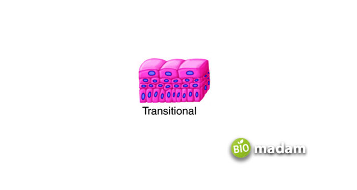
Stratified Transitional Epithelium

It is another sub-type of complex, stratified epithelial cells, categorized based on cell layers. Such tissues are multi-layered, also are urothelium, and cannot change their shape or composition. They are located in the urinary tract, including the renal pelvis, urethra, urinary bladder, and ureters.
This epithelium becomes less stratified (flatten) and transits from cuboidal to squamous shape whenever the bladder gets filled with urine. But soon as the urine passes out, the cells return to the cuboidal shape with a domed apex (non-stretched) form.
Glandular Epithelial Tissues
The third and last type of epithelial tissue is the glandular tissue which is the most modified form. These cells function to get blood substances and alter them to release in the respective cell. Glandular tissues are found dispersed in the epithelial covering as unicellular structures or in glandular organs. For example, thyroid glands and goblet cells in the intestinal epithelium.
These tissues are further classified based on the point of product release and the number of cells involved. These are:
- Endocrine and Exocrine Glands
- Unicellular Glands
- Multicellular Glands
Conclusion
The epithelial tissues are as important for a body as any other organ. The proper functioning of all types of epithelial tissue helps a body regulate its mechanisms, such as osmosis, filtration, secretion, etc. Sometimes the genetic disorder in these epithelial tissues leads to severe lifetime infections or diseases, for example, ichthyosis.

Jeannie has achieved her Master’s degree in science and technology and is further pursuing a Ph.D. She desires to provide you the validated knowledge about science, technology, and the environment through writing articles.

