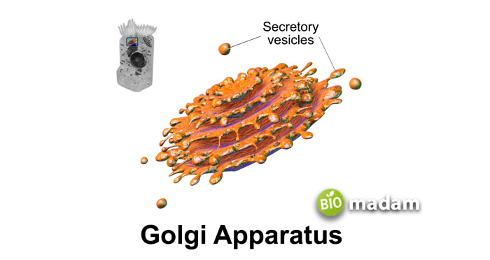
Golgi bodies are the organelles present in all eukaryotic cell. However, they are not the same. Golgi bodies or Golgi apparatus are protein and lipid metabolizing and transportation sites in the cell. They are present near the nucleus, extending towards the endoplasmic reticulum and plasma membrane.
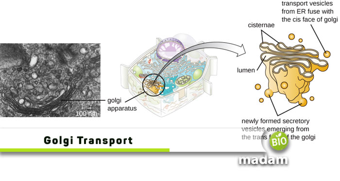
The Golgi bodies look like a stack of multiple layers or discs adjusted over each other. Cells typically have 4-10 folds or cisternae in each structure. Some organisms may have more cisterna in each Golgi apparatus. One side of the Golgi apparatus is known as the cis side, while the other is the trans side. The area between these two is known as the medial. Animal cells contain only one Golgi body, while plants may have multiple.
Despite the small size, the Golgi apparatus performs various important functions within the cell. It is involved in protein glycosylation, and lipid and polysaccharide metabolism. Protein transportation is one of the main functions of Golgi bodies. The Golgi apparatus sorts and assembles several types of proteins before encasing them into adequate vesicles to be transported to other areas.
Let’s tell you how protein transport in the Golgi bodies takes place.
Arrival of Proteins
The endoplasmic reticulum, which comprises smooth and rough endoplasmic reticulum, produces proteins and sends them to the Golgi apparatus. The proteins enter the assembly through the cis side, which faces the endoplasmic reticulum. The proteins keep moving through the various layers of the organelle and go through different processes. These processes prepare them for transport to parts of the cell or outside the cell per need. Some proteins are also stored by the Golgi bodies that they require for adequate functioning.
Modification
The cisternae of Golgi bodies contain enzymes that facilitate the modification of specific enzymes. They add or remove carbohydrates from proteins to carry out processes like glycosylation. These enzymes may also contribute to phosphorylation or sulfation within the Golgi bodies. Enzymes called “resident enzymes” help modify cargo proteins in each cisterna.
Protein Transport
The movement and transport of protein can be explained through two models:
- Vesicular transport model
- Cisternal maturation model
Vesicular Transport Model
The vesicular transport model explains the transport of proteins through vesicles as they were initially identified to be associated with the Golgi apparatus. George Palade and Marilyn Farquhar developed this model. It states that the vesicles fuse with the cisternae membrane to move from one cisterna to the other. Thus, it also endorsed the theory that these cisternae contain specific enzymes that help modify the proteins by adding or removing sulfate groups or sugars. It is also involved in transporting molecules back to the endoplasmic reticulum. This model emphasizes that cisternae do not move.

Cisternal Maturation Model
The cisternal maturation model explains the Golgi bodies as a dynamic organelle compared to the vesicular transport model. Opposed to the vesicular transport model, this model does not mention that the cisternae are static. Instead, the cisternal maturation model mentions that the cis cisternae eventually mature into trans cisternae. New cisternae keep developing at the cis side. According to this model, the vesicles do not modify or transport proteins in the Golgi apparatus. The vesicles are only used to transport material back to the endoplasmic reticulum.

Vesicular Transport Model vs. Cisternal Maturation Model
The presence of multiple models regarding the transport of proteins within the Golgi apparatus piqued the curiosity of researchers. More studies were conducted to understand the exact mechanism of protein transport in Golgi bodies. Recently, in the 1990s, researchers studied different cells and tissues to understand the exact mechanism of Golgi bodies. They examined mammalian cells through immunoelectron microscopes. The observations showed procollagens in the cisternae but not in the vesicles. Another team of scientists observed protists with flagella that developed and transported scale-like material. However, again, these scales were not found in vesicles.
Thus, it provided evidence in favor of the cisternal maturation model, which advocates the maturation of the cis side into the trans side. It was further vetted by a visual obtained by live-cell fluorescence microscopy. The microscope observation showed cisternal maturation in the Golgi apparatus in yeast. It is easier to observe this process in Saccharomyces cerevisiae as they have a comparatively scattered structure. The cisternae appear irregularly dispersed individually within the cell. So, this method gave a better insight into the protein transport and Golgi maturation phenomenon.
The process appeared as changes in the color of the protein molecule as a result of cisternal maturation observed by fluorescent microscopy.
It shows that the observations support the cisternal maturation model as the cisternae mature; thus, the proteins change their color over time. It exhibits a change in the composition of different types of proteins within Golgi bodies. On the other hand, a continuous movement of proteins in the same color (in vesicles) would have supported the vesicular transport model. The research also showed a gradual change in the exact shape of the Golgi body’s structure as a result of maturation.
The Bottom Line
Golgi bodies are involved in modifying and transporting proteins and lipids within the cell and outside. The materials, such as proteins, enter the Golgi apparatus through the cis side and move towards the trans side to exit. During their movement, the enzymes in the cisternae modify the proteins. The initial vesicular transport model mentioned that these proteins move within vesicles to the other side, and the cisternae are static. Yet, another research suggested that the cisternae are more dynamic and mature over time; the cis face of the Golgi apparatus continuously matures and forms the trans face. Recent studies support the cisternal maturation model as the researchers observed changes in the structure of the Golgi apparatus and protein composition.

Hello, I would like to introduce myself to you! I am Chelsea Rogers, an experienced blog writer for science articles, holding an MPhil degree. My enthusiasm to grab the best knowledge, let it relate to botany, zoology, or any other science branch. Read my articles & let me wait for your words s in the comment section.

